. 2004 Jan;113(1):28-37.
doi: 10.1172/JCI19491.

Stat-3 is required for pulmonary homeostasis during hyperoxia

Affiliations

Stat-3 is required for pulmonary homeostasis during hyperoxia

Isamu Hokuto et al. J Clin Invest. 2004 Jan.

Abstract

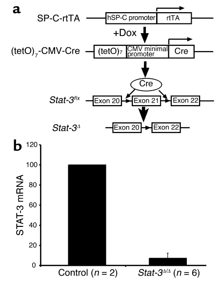
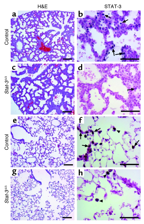
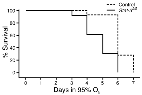
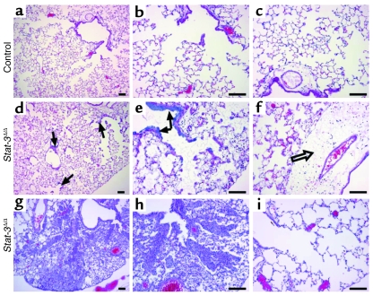
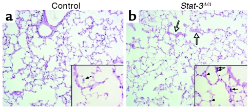
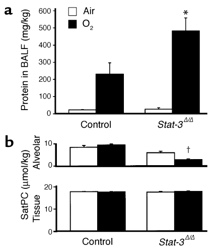
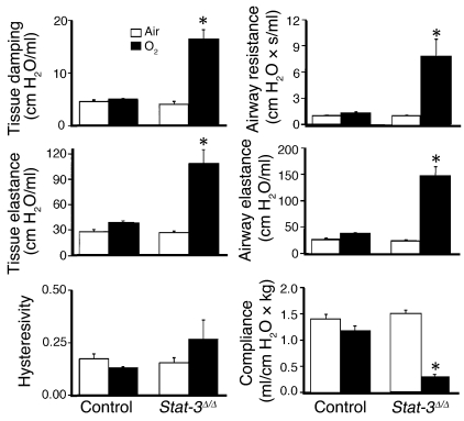
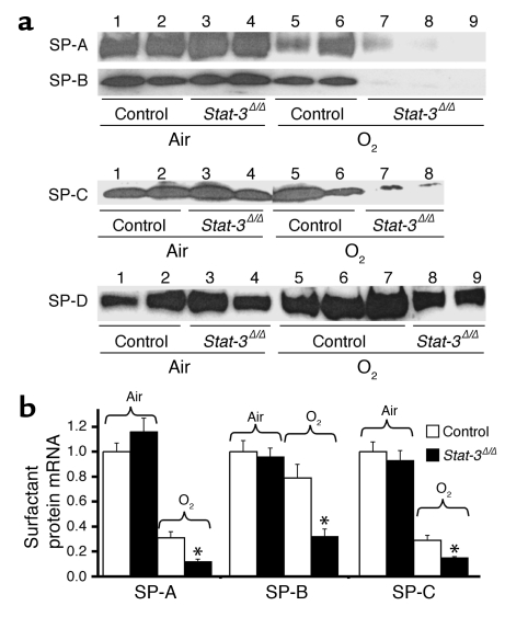
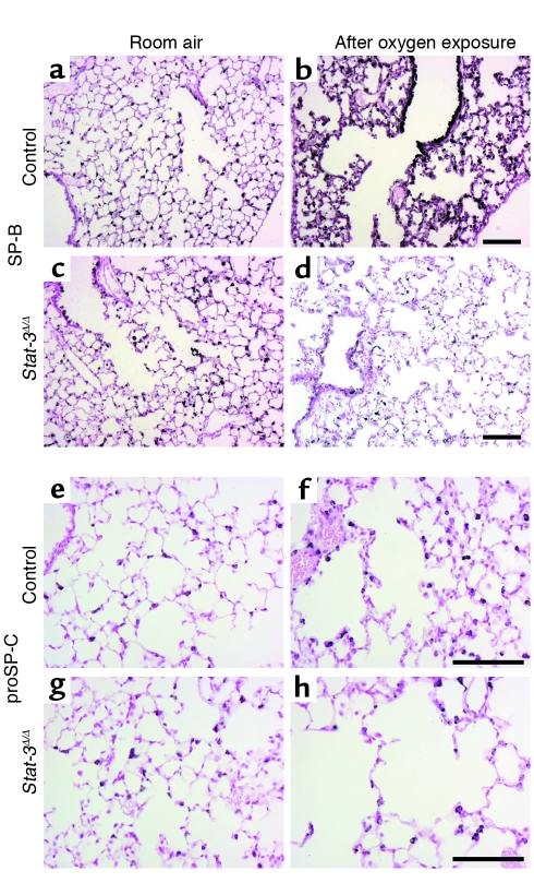
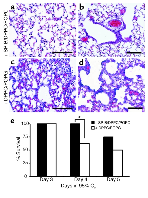
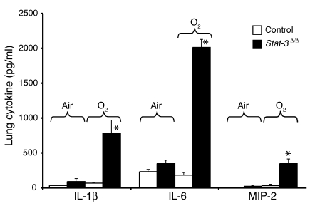
Acute lung injury syndromes remain common causes of morbidity and mortality in adults and children. Cellular and physiologic mechanisms maintaining pulmonary homeostasis during lung injury remain poorly understood. In the present study, the Stat-3 gene was selectively deleted in respiratory epithelial cells by conditional expression of Cre-recombinase under control of the surfactant protein C gene promoter. Cell-selective deletion of Stat-3 in respiratory epithelial cells did not alter prenatal lung morphogenesis or postnatal lung function. However, exposure of adult Stat-3-deleted mice to 95% oxygen caused a more rapidly progressive lung injury associated with alveolar capillary leak and acute respiratory distress. Epithelial cell injury and inflammatory responses were increased in the Stat-3-deleted mice. Surfactant proteins and lipids were decreased or absent in alveolar lavage material. Intratracheal treatment with exogenous surfactant protein B improved survival and lung histology in Stat-3-deleted mice during hyperoxia. Expression of Stat-3 in respiratory epithelial cells is not required for lung formation, but plays a critical role in maintenance of surfactant homeostasis and lung function during oxygen injury.


Figures

Figure 1
Figure 1
Conditional deletion of Stat-3 in respiratory epithelium. (a) The human SP-C (hSP-C) promoter was used to express the reverse tetracycline transactivator (rtTA). In the presence of doxycycline (Dox), rtTA binds to the (tetO)7 promoter, activating transcription of Cre-recombinase. The loxP sites were inserted in introns 20 and 21 of the Stat-3 gene (Stat-3flx), deleting exon 21 after recombination, to produce the Stat-3Δ locus. (b) STAT-3 RNA was assessed in purified alveolar type II cells. RNA was extracted from primary cultures of alveolar type II cells isolated from Stat-3Δ/Δ and control mice. RNA from control (n = 2) or Stat-3Δ/Δ (n = 2 or 4) were pooled and analyzed by real-time RT-PCR. Data were standardized to β-actin RNA and normalized to control. In the Stat-3Δ/Δ cells, STAT-3 mRNA was less than 10% of control values.
Figure 2
Figure 2
STAT-3 immunostaining in the Stat-3Δ/Δ mice. Dams were treated with doxycycline from E0. Lung sections were prepared at E18.5 (ad) or at 6 weeks of age (eh). Tissues were stained with H&E (a, c, e, g) or immunostained for STAT-3 (b, d, f, h). Lung morphology was not altered in the Stat-3Δ/Δ mice. Complete or nearly complete loss of STAT-3 staining was observed in respiratory epithelial cells at E18. Likewise, staining was observed in alveolar type II and conducting airway cells at PN 6 weeks in Stat-3Δ/Δ mice (arrows). STAT-3 was detected in alveolar macrophages and in other nonepithelial cells in control and Stat-3Δ/Δ mice (f and h) (arrowheads). Figures are representative of n ≥ 3 per genotype. Scale bar: 100 μm (a, c, e, g) or 50 μm (b, d, f, h).
Figure 3
Figure 3
Kaplan-Meier plot of survival of Stat-3Δ/Δ and control mice during hyperoxia. Adult Stat-3Δ/Δ mice and littermates were exposed to 95% O2. Survival of Stat-3Δ/Δ mice in hyperoxia (n = 13) was significantly decreased compared with littermate, nondeleted controls (n = 14). P < 0.001 by log-rank test.
Figure 4
Figure 4
Lung morphology after exposure to 95% FiO2. Adult (8–10 weeks of age) Stat-3Δ/Δ mice and nondeleted littermates were exposed to 95% FiO2 for 65 hours. Minor histologic changes (perivascular edema) in lung histology were observed in control mice after oxygen exposure (ac). Severe epithelial cell necrosis (arrows), thickened alveoli, lymphotic edema (open arrow), and extensive inflammation were observed in the Stat-3Δ/Δ mice (df). Airspace enlargement was observed in some Stat-3Δ/Δ mice (gi). Photomicrographs are representative of n = 7 in each group. Scale bar: 100 μm.
Figure 5
Figure 5
STAT-3 immunohistochemistry after exposure to 95% oxygen. Lungs from control (a) and Stat-3Δ/Δ (b) mice were stained for Stat-3 after 72 hours of exposure to 95% O2. Alveolar type II cells, bronchiolar epithelial cells, and alveolar macrophages were stained in controls. Stat-3 was detected in inflammatory cells (filled arrows), vascular and stromal cells, but not in epithelial cells (arrowheads), in Stat-3Δ/Δ mice. Bronchiolar epithelial cell necrosis was prominent (open arrows). Original magnification ×10. Magnification for inserted enlarged photomicrographs ×40.
Figure 6
Figure 6
Increased alveolar capillary leak and decreased surfactant in Stat-3Δ/Δ mice after hyperoxia. (a) Total protein concentration was measured in lung lavage fluid 65 hours after exposure to 95% O2. Protein content was similar in control and Stat-3Δ/Δ mice in room air. After oxygen exposure, BALF protein recovered from each mouse (micrograms per kilogram) was significantly increased in the Stat-3Δ/Δ mice, n = 5 per group. *P < 0.05 versus others and P < 0.001 versus room air, as assessed by ANOVA. (b) Surfactant saturated phosphatidylcholine (SatPC) was significantly decreased in BALF from the Stat-3Δ/Δ mice following hyperoxia. Statistical differences were analyzed by ANOVA. P < 0.001 versus air.
Figure 7
Figure 7
Abnormalities in pulmonary mechanics in Stat-3Δ/Δ mice following hyperoxia. Lung mechanics were assessed in adult control and Stat-3Δ/Δ mice exposed to room air or 95% O2 for 65 hours. Lung mechanics were similar in control and Stat-3Δ/Δ mice in room air. After exposure to 95% O2, airway resistance, airway elastance, tissue damping, and tissue elastance were increased and compliance decreased significantly in Stat-3Δ/Δ but not in control mice. n = 5 per group. Statistical differences were analyzed by ANOVA. *P < 0.01 versus control group.
Figure 8
Figure 8
Decreased surfactant proteins in Stat-3Δ/Δ mice following hyperoxia. (a) SP-A, SP-B, SP-C, and SP-D were quantitated in BAL fluid by Western blot analysis. In room air, SP-A, SP-B, SP-C, and SP-D were not altered (lanes 3 and 4). After exposure to 95% O2 for 65 hours, SP-A and SP-B were significantly decreased in the Stat-3Δ/Δ mice (lanes 7–9), P < 0.01, after quantitative imaging. (b) SP-A, SP-B, and SP-C mRNAs were quantitated by S1 nuclease assay. In room air, SP-A, SP-B, and SP-C mRNAs were similar in Stat-3Δ/Δ and control mice. After exposure to 95% O2 for 65 hours, SP-A, SP-B, and SP-C mRNAs were decreased in Stat-3Δ/Δ compared with control mice. Results were standardized to L32 mRNA and normalized to room air controls, n = 5 per group. Statistical differences were assessed by Student’s t test, *P < 0.01 versus control O2.
Figure 9
Figure 9
Decreased immunostaining for SP-B and proSP-C in Stat-3Δ/Δ mice after exposure to 95% FiO2. Staining for SP-B (ad) and proSP-C (eh) was performed in Stat-3Δ/Δ and control littermates exposed to room air or 95% O2. In room air there were no differences in intensity or distribution of SP-B and proSP-C staining between Stat-3Δ/Δ and control mice. After 65 hours in 95% oxygen, SP-B and proSP-C staining were decreased in Stat-3Δ/Δ (d and h) compared with controls (b and f). Scale bar: 100 μm. Photomicrographs are representative of n ≥ 3.
Figure 10
Figure 10
Exogenous SP-B protects Stat-3Δ/Δ mice during hyperoxia. (a) Lung morphology of the Stat-3Δ/Δ mice treated with SP-B/DPPC/POPG (a and b) or DPPC/POPG (c and d) during hyperoxia exposure is shown. Lungs were excised 65 hours after exposure to 95% oxygen and stained with H&E. The lungs of SP-B/DPPC/DOPG–treated mice had less inflammatory cell infiltration, and epithelial cell necrosis was not observed (a and b). Alveolar thickening, inflammation, and epithelial cell necrosis was widespread in mice treated with DPPC/DOPG alone (c and d). Photomicrographs are representative of n = 5 from each group. Bars = 100 μm. (e) SP-B enhances survival of Stat-3Δ/Δ mice during hyperoxia. Stat-3Δ/Δ mice were placed in 95% oxygen and treated (intratracheally) with SP-B/DPPC/DOPG or DPPC/DOPG as described in Methods. Survival on day 4 was significantly increased in SP-B/DPPC/DOPG–treated mice; *P < 0.05. On day 5, more SP-B/DPPC/DOPG–treated mice survived, but differences were not statistically significant; n = 8 per group.
Figure 11
Figure 11
Increased IL-1β, IL-6, and MIP-2 in Stat-3Δ/Δ mice after 95% O2. IL-1β, IL-6, and MIP-2 were measured in lung tissue from control and Stat-3Δ/Δ mice exposed to room air or 95% O2 for 65 hours. Levels of each cytokine were significantly increased in the Stat-3Δ/Δ mice after hyperoxia, n = 5 per group, as determined by ANOVA; *P < 0.001 versus others.

Similar articles

Cited by

References

    1. Akira S. IL-6-regulated transcription factors. Int. J. Biochem. Cell Biol. 1997;29:1401–1418. - PubMed
    1. Darnell JE., Jr STATs and gene regulation. Science. 1997;277:1630–1635. - PubMed
    1. Boccaccio C, et al. Induction of epithelial tubules by growth factor HGF depends on the STAT pathway. Nature. 1998;391:285–288. - PubMed
    1. Takeda K, et al. Targeted disruption of the mouse Stat3 gene leads to early embryonic lethality. Proc. Natl. Acad. Sci. U. S. A. 1997;94:3801–3804. - PMC - PubMed
    1. Levy DE, Lee CK. What does Stat3 do? J. Clin. Invest. 2002;109:1143–1148. doi:10.1172/JCI200215650. - PMC - PubMed

Publication types

MeSH terms