. 2004 Jan;113(1):74-84.
doi: 10.1172/JCI17005.

Cure of prediabetic mice by viral infections involves lymphocyte recruitment along an IP-10 gradient

Affiliations

Cure of prediabetic mice by viral infections involves lymphocyte recruitment along an IP-10 gradient

Urs Christen et al. J Clin Invest. 2004 Jan.

Abstract

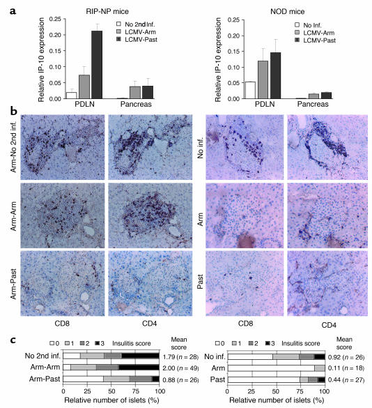
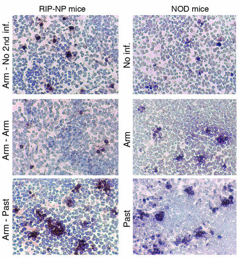
Viruses can cause but can also prevent autoimmune disease. This dualism has certainly hampered attempts to establish a causal relationship between viral infections and type 1 diabetes (T1D). To develop a better mechanistic understanding of how viruses can influence the development of autoimmune disease, we exposed prediabetic mice to various viral infections. We used the well-established NOD and transgenic RIP-LCMV models of autoimmune diabetes. In both cases, infection with the lymphocytic choriomeningitis virus (LCMV) completely abrogated the diabetic process. Interestingly, such therapeutic viral infections resulted in a rapid recruitment of T lymphocytes from the islet infiltrate to the pancreatic draining lymph node, where increased apoptosis was occurring. In both models this was associated with a selective and extensive expression of the chemokine IP-10 (CXCL10), which predominantly attracts activated T lymphocytes, in the pancreatic draining lymph node, and in RIP-LCMV mice it depended on the viral antigenic load. In RIP-LCMV mice, blockade of TNF-alpha or IFN-gamma in vivo abolished the prevention of T1D. Thus, virally induced proinflammatory cytokines and chemokines can influence the ongoing autoaggressive process beneficially at the preclinical stage, if produced at the correct location, time, and levels.


Figures

Figure 1
Figure 1
Prevention of T1D in NOD and RIP-LCMV mice by LCMV infection given during the prediabetic phase. (a) Cumulative incidence of diabetes in RIP-LCMV-NP mice infected i.p. with 105 PFUs of LCMV-Arm on day 0. One month after infection, mice were infected i.p. with 105 PFUs of either LCMV-Arm (Arm-Arm) or LCMV-Past (Arm-Past). Blood glucose was measured in biweekly intervals for at least 9 months, and values greater than 300 mg/dl were considered diabetic. The number of diabetic mice and the total number of animals used per group are indicated in brackets. (b) Groups of 10–15 RIP-LCMV-NP H-2b mice were infected i.p. with 105 PFUs of various LCMV strains (single infection). Blood glucose values were determined as described in Methods. The NP escape variant contains a single amino acid change from F to L in position AA404 of the immunodominant H-2Db NP396 peptide, which prevents binding of the variant NP396 to H-2Db (36). pCTL analysis was performed 7 days after infection as described in Methods. Note that RIP-NP mice express the LCMV-NP antigen in their thymus in addition to their islets and therefore have reduced numbers of NP CTLs in their periphery and develop diabetes more slowly, as reported by us previously (24). Non-Tg, nontransgenic C57BL/6 mice. (c) Groups of 10–15 NOD mice were infected i.p. with 105 PFUs of LCMV-Arm (single infection) at week 10 or 20 of age. Blood glucose values were determined every 4 weeks.
Figure 2
Figure 2
Protective viral infection results in increased IP-10 expression in the PDLN and a decrease of islet-infiltrating CD8 lymphocytes. Left: RIP-LCMV-NP mice were infected (inf.) i.p. with 105 PFUs of LCMV-Arm and received a secondary i.p. infection (2nd inf.) after 1 month with 105 PFUs of LCMV-Arm or LCMV-Past. Right: Nine-week-old NOD mice were infected i.p. with 105 PFUs of LCMV-Arm (Arm) or LCMV-Past (Past). (a) Some RIP-LCMV-NP and NOD mice were sacrificed at day 1 after secondary infection for assessment of the expression of IP-10 (CXCR3) in the pancreas and PDLN. The relative IP-10 mRNA expression was analyzed by RNase protection assay and normalized against L32 housekeeping RNA expression (n = 3–4). (b) At day 3 after secondary infection, pancreata and PDLNs were harvested, and 6-μm tissue sections were probed for cellular infiltration by CD4 and CD8 T cells. These representative tissue sections show an average degree of islet infiltration per group as found in the pancreas of individual mice (n = 3–4). (c) Insulitis score as obtained from sections of 3–4 mice per group. Scoring system: 0, no infiltration; 1, some peri-insular infiltration; 2, heavy peri-insular infiltration with some intra-insular infiltrates; 3, heavy intra-insular infiltration and/or islet scars. The mean score was obtained by division of the sum of all individual islet-infiltration scores by the total number of islets analyzed.
Figure 3
Figure 3
Abrogation of diabetes following LCMV infection is associated with increased apoptosis in the PDLN of NOD and RIP-LCMV mice. RIP-LCMV-NP mice were infected i.p. with 105 PFUs of LCMV-Arm and received a secondary i.p. infection after 1 month with 105 PFUs of LCMV-Arm or LCMV-Past. Nine-week-old NOD mice were infected i.p. with 105 PFUs of LCMV-Arm or LCMV-Past. At day 3 after infection, PDLNs were harvested, and 6-μm tissue sections were probed for apoptotic cells using TUNEL staining. The staining shown represents an average number of apoptotic cells per group as found in individual mice (n = 3–4). Note the formation of clusters of apoptotic cells after infection with LCMV-Past in both RIP-NP and NOD mice.
Figure 4
Figure 4
In RIP-LCMV mice, apoptosis of autoaggressive (LCMV-specific) CD8 lymphocytes in the PDLN is enhanced after protective viral infection. (a) Frequency of LCMV-NP–specific pCTLs in RIP-LCMV-NP mice after secondary i.p. infection with 105 PFUs of LCMV-Arm or LCMV-Past administered 4 weeks after initial LCMV-Arm infection. Note that the reduced pCTL frequency in the PDLN after secondary infection with LCMV-Past correlated with the drastically lower incidence of diabetes in those mice. *pCTL frequencies in the spleen at day 3 after primary i.p. infection with 105 PFUs of LCMV-Arm or LCMV-Past are displayed as a reference. nd, not determined. (b) RIP-LCMV-NP mice were infected i.p. with 105 PFUs of LCMV-Arm and, after 1 month, with 105 PFUs of either LCMV-Arm or LCMV-Past. At day 3 after secondary infection, PDLN CD8 lymphocytes were stained with anti-CD8 mAb, H-2Db(GP33)- or H-2Db(NP396)-tetramers, and annexin V. CD8hi, H-2Db-tetramerhi cells (left panels, dot blots) were analyzed with annexin V for apoptosis (right panels, histograms). The relative numbers of CD8hi, H-2Db-tetramerhi cells and annexin Vhi cells are indicated as percentage of gated cells.
Figure 5
Figure 5
Abrogative infection of RIP-LCMV mice with LCMV-Past results in decreased pCTL frequencies and higher viral titers in the PDLN. Viral titers were determined in the pancreas, the spleen, and the PDLN of RIP-LCMV-NP mice at day 3 after primary i.p. infection with 105 PFUs of LCMV-Arm or LCMV-Past or, alternatively, at day 3 after secondary infection (administered 4 weeks after initial LCMV-Arm infection) with 105 PFUs of LCMV-Arm (nonabrogative) or LCMV-Past (abrogates diabetes). Data are shown as mean PFUs/10 mg tissue (± SD) as obtained from plaque assays (n = 4).
Figure 6
Figure 6
Blockade of IFN-γ or TNF-α prevents abrogation of diabetes by secondary viral infection. (a) Cumulative incidence of diabetes in RIP-LCMV-NP mice infected i.p. with 105 PFUs of LCMV-Arm on day 0 and 105 PFUs of LCMV-Past 1 month after LCMV-Arm infection. Blood glucose was measured in weekly intervals, and values greater than 300 mg/dl were considered diabetic. The number of diabetic mice and the total number of animals used per group are indicated in brackets. Human TNFR55-IgG1 fusion protein (38, 39) or neutralizing rat anti–mouse IFN-γ antibody (Pharmingen, San Diego, California, USA) were given at 100 μg (i.v.) or 50 μg (i.v.), respectively, at days 1, 3, 6, and 9 after secondary (Past) infection. (b) In vitro hyperstimulation with Db(NP396)-tetramers (16 hours at 37°C) of lymphocytes isolated from spleen and PDLN of C57BL/6 mice at 1 week after LCMV-Arm infection. Apoptosis of Db(NP396)-specific CD8 T cells was determined by staining with annexin V–phycoerythrin conjugate in parallel to staining with Db(NP396)-tetramers. Cells were incubated in medium alone (control) or in the presence of human TNFR55-IgG1 fusion protein (100 μg/well) or neutralizing rat anti–mouse IFN-γ antibody (100 μg/well). *Among splenocytes, significant differences in the number of Db(NP396)-specific CD8 T cells that undergo apoptosis (annexin Vhi cells) were observed after blockade of TNF-α compared with the control stimulation, but not after IFN-γ blockade (Student’s t test, P < 0.05). **PDLNs were collected from three mice, and cells were pooled in order to increase the number of Db(NP396)-specific CD8 T cells within the experimental setup.

References

    1. Redondo MJ, et al. Genetic determination of islet cell autoimmunity in monozygotic twin, dizygotic twin, and non-twin siblings of patients with type 1 diabetes: prospective twin study. BMJ. 1999;318:698–702. - PMC - PubMed
    1. Kallmann BA, et al. Cytokine secretion patterns in twins discordant for Type I diabetes. Diabetologia. 1999;42:1080–1085. - PubMed
    1. Thomson G, et al. Genetic heterogeneity, modes of inheritance, and risk estimates for a joint study of Caucasians with insulin-dependent diabetes mellitus. Am. J. Hum. Genet. 1988;43:799–816. - PMC - PubMed
    1. Honeyman MC, et al. Association between rotavirus infection and pancreatic islet autoimmunity in children at risk of developing type 1 diabetes. Diabetes. 2000;49:1319–1324. - PubMed
    1. Notkins, A.L., and Yoon, J.-W. 1984. Virus-induced diabetes mellitus. In Concepts in viral pathogenesis. A.L. Notkins and M.B.A. Oldstone, editors. Springer-Verlag. New York, New York, USA. 241–247.

Publication types

MeSH terms