

Review
doi: 10.1039/b303439a.
Demystifying the three dimensional structure of G protein-coupled receptors (GPCRs) with the aid of molecular modeling
Affiliations
- PMID: 14703805
- PMCID: PMC9388203
- DOI: 10.1039/b303439a
Item in Clipboard
Review
Demystifying the three dimensional structure of G protein-coupled receptors (GPCRs) with the aid of molecular modeling
Chem Commun (Camb).
.
Abstract
We review our recent work on adenosine receptors, a family of GPCRs; focusing our attention on A3 adenosine receptor, we have demonstrated that the reciprocal integration of different theoretical and experimental disciplines can be very useful for the successful protein-based design of new, potent and selective receptor ligands.
Figures
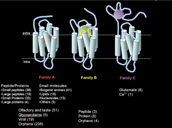
The “world of human GPCRs”. A distribution of the 508 human GPCRs that have been discovered so far, grouped according to the type of natural ligand that binds to them. Orphan receptors are receptors of yet unknown function.
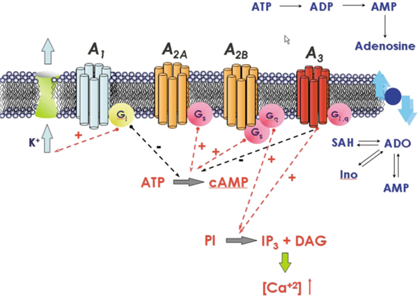
Signal transduction pathways associate with the activation of the human adenosine receptors.
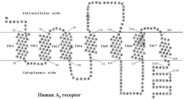
Heptahelical diagram of the human A3 adenosine receptor. The putative transmembrane domains were modified according to the high resolution rhodopsin model. Amino acids mutated in the present study are circled. Residues 286–295 correspond to an extra helical domain in rhodopsin, which is discontinuous from TM7.
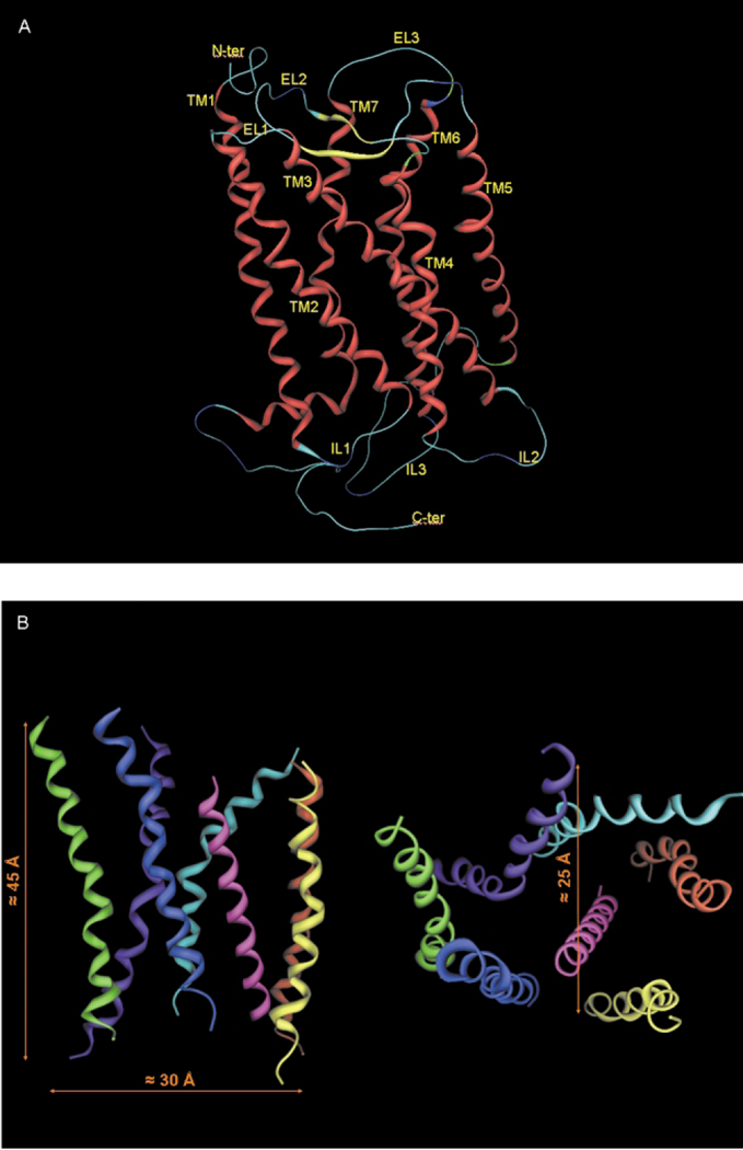
A) Stereoview of the complete topology of human A3 receptor obtained by a homology modeling approach. B) Stereoview of the human A3 receptor transmembrane helical bundle model viewed along the helical axes from the extracellular end (right) and perpendicular to the helical axes (left).
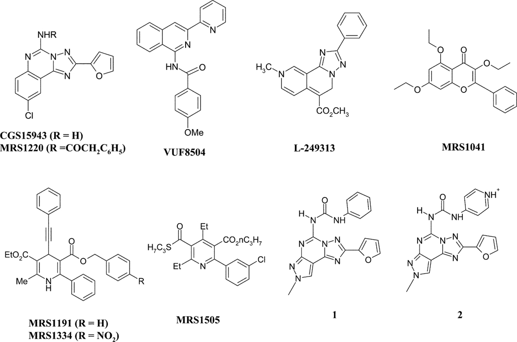
Chemical structures of the most representative members of the several classes of heterocyclic compounds identified as A3 adenosine antagonists.
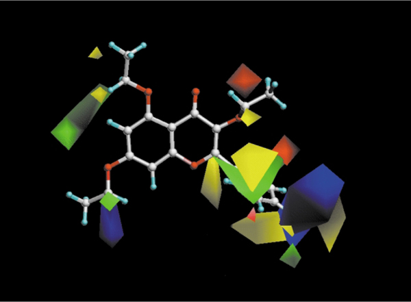
CoMFA steric and electrostatic contour plots from the analysis based on the A3 receptor 3D-QSAR without cross-validation. Compound MRS1041 shown inside the field. Favoring activity: green, bulky group (contribution level 80%); yellow, less bulky; blue, positive charge (contribution level 70%); red, negative charge.
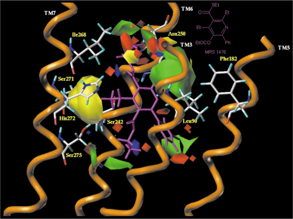
Side view of the human A3–MRS1476 complex model. The side chains of the important residues in proximity ( < 5 Å) to the docked pyridine molecule are highlighted and labeled: Leu90 (TM3), Phe182 (TM5), Ser242 (TM6), Ser247 (TM6), Asn250 (TM6), Ser271 (TM7), His272 (TM7) and Ser275 (TM7). The steric and the electrostatic contour plots, obtained from the CoMFA analysis, are included into the ligand binding cavity.
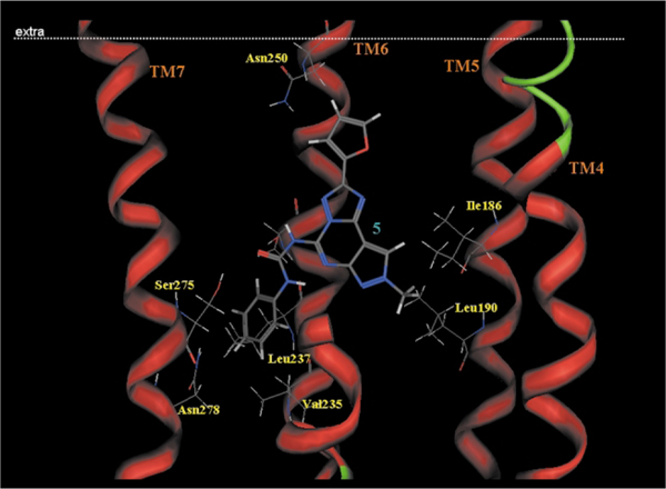
Side view of the human A3–5 complex model. The side chains of the important residues in proximity to the docked 5 molecule are highlighted and labeled.
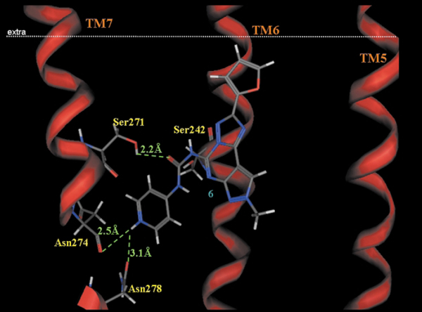
Side view of the human A3–6 complex model. The side chains of the important residues in proximity to the docked 6 molecule are highlighted and labeled.
Similar articles
-
Molecular modeling the human A1 adenosine receptor and study of the mechanisms of its selective ligand binding.Dokl Biochem Biophys. 2002 Sep-Oct;386:271-4. doi: 10.1023/a:1020715812906. Dokl Biochem Biophys. 2002. PMID: 12469507 No abstract available.
-
Limits of ligand selectivity from docking to models: in silico screening for A(1) adenosine receptor antagonists.PLoS One. 2012;7(11):e49910. doi: 10.1371/journal.pone.0049910. Epub 2012 Nov 21. PLoS One. 2012. PMID: 23185482 Free PMC article.
-
Structure and function of adenosine receptor heteromers.Cell Mol Life Sci. 2021 Apr;78(8):3957-3968. doi: 10.1007/s00018-021-03761-6. Epub 2021 Feb 12. Cell Mol Life Sci. 2021. PMID: 33580270 Free PMC article. Review.
-
Structural Mapping of Adenosine Receptor Mutations: Ligand Binding and Signaling Mechanisms.Trends Pharmacol Sci. 2018 Jan;39(1):75-89. doi: 10.1016/j.tips.2017.11.001. Epub 2017 Dec 5. Trends Pharmacol Sci. 2018. PMID: 29203139 Review.
-
Nomenclature and classification of purinoceptors.Pharmacol Rev. 1994 Jun;46(2):143-56. Pharmacol Rev. 1994. PMID: 7938164 Free PMC article. Review. No abstract available.
Cited by
-
The Neuroprotective Effects of Moderate and Regular Caffeine Consumption in Alzheimer's Disease.Oxid Med Cell Longev. 2021 Aug 17;2021:5568011. doi: 10.1155/2021/5568011. eCollection 2021. Oxid Med Cell Longev. 2021. PMID: 34447487 Free PMC article. Review.
-
Prediction of the 3-D structure of rat MrgA G protein-coupled receptor and identification of its binding site.J Mol Graph Model. 2007 Nov;26(4):800-12. doi: 10.1016/j.jmgm.2007.07.003. Epub 2007 Jul 14. J Mol Graph Model. 2007. PMID: 17728165 Free PMC article.
-
Progress in the pursuit of therapeutic adenosine receptor antagonists.Med Res Rev. 2006 Mar;26(2):131-59. doi: 10.1002/med.20048. Med Res Rev. 2006. PMID: 16380972 Free PMC article. Review.
-
Forced unbinding of GPR17 ligands from wild type and R255I mutant receptor models through a computational approach.BMC Struct Biol. 2010 Mar 16;10:8. doi: 10.1186/1472-6807-10-8. BMC Struct Biol. 2010. PMID: 20233425 Free PMC article.
-
Pyrazolo-triazolo-pyrimidines as adenosine receptor antagonists: Effect of the N-5 bond type on the affinity and selectivity at the four adenosine receptor subtypes.Purinergic Signal. 2008 Mar;4(1):39-46. doi: 10.1007/s11302-007-9058-y. Epub 2007 Jul 25. Purinergic Signal. 2008. PMID: 18368532 Free PMC article.
References
-
- Wilson S. and Bergsma D, Drug Des. Discov, 2000, 17, 105–114. - PubMed
-
- Civelli O, Nothacker HP, Saito Y, Wang Z, Lin SH and Reinscheid RK, Trends Neurosci. , 2001, 24, 230–237. - PubMed
-
- Marchese A, George SR, Kolakowski LF Jr, Lynch KR and O’Dowd BF, Trends Pharmacol. Sci, 1999, 20, 370–375. - PubMed
-
- Lee DK, George SR, Evans JF, Lynch KR and O’Dowd BF, Curr. Opin. Pharmacol, 2001, 1, 31–39. - PubMed
Publication types
MeSH terms
Substances
Grants and funding
LinkOut - more resources
Full Text Sources
Other Literature Sources