. 2004 Jan 13;101(2):552-7.
doi: 10.1073/pnas.0307437100. Epub 2004 Jan 2.

Conditional targeting of E-cadherin in skin: insights into hyperproliferative and degenerative responses

Affiliations

Conditional targeting of E-cadherin in skin: insights into hyperproliferative and degenerative responses

Christopher L Tinkle et al. Proc Natl Acad Sci U S A. .

Abstract

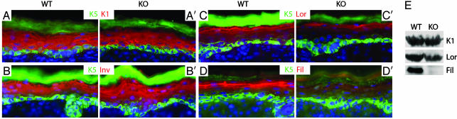
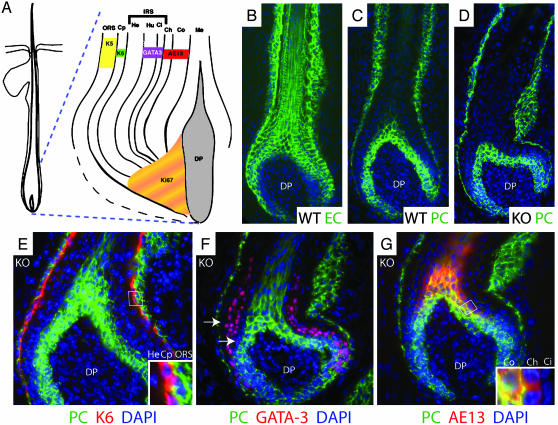
Loss of E-cadherin has been associated with human cancers, and yet in the early mouse embryo and the lactating mammary gland, the E-cadherin null state results in tissue dysfunction and cell death. Here we targeted loss of E-cadherin in skin epithelium. The epidermal basal layer responded by elevating P-cadherin, enabling these cells to maintain adherens junctions. Suprabasal layers upregulated desmosomal cadherins, but without classical cadherins, terminal differentiation was impaired. Progressive hyperplasia developed with age, a possible consequence of proliferative maintenance in basal cells coupled with defects in terminal differentiation. In contrast, hair follicles lost integrity of the inner root sheath and hair cuticle without apparent elevation of cadherins. These findings suggest that, if no compensatory mechanisms exist, E-cadherin loss may be incompatible with epithelial tissue survival, whereas partial compensation can result in alterations in differentiation and proliferation.


Figures

Fig. 1.
Fig. 1.
Generation of mice conditionally null for E-cadherin in skin epithelium. (A) Animals whose epidermis is either heterozygous or homozygous for the WT E-cadherin allele are referred to as WT; those whose epidermis is homozygous for the K14-Cre-recombined mutant E-cadherin allele are called KO. Shown are newborn pups. (B) PCR confirmation of genotype (Top and Middle) and anti-E-cadherin (EC) Western blot of epidermal proteins (Bottom). (C and C′) Immunofluorescence of skin sections labeled with Abs indicated (color coding is according to secondary antibodies). * denotes nonspecific staining of cornified layer with the secondary FITC-conjugated Ab. (D–E′) Toluidine blue-stained semithin sections (1 μm) of newborn backskin. BL, basal layer; SL, spinous layer; GL, granular layer; CL, cornified layer. (E and E′ are 5× the magnification of D and D′.) Arrows denote regions where intercellular spaces are widened. epi, epidermis, de, dermis; hf, hair follicle.
Fig. 2.
Fig. 2.
Up-regulation of P-cadherin in basal but not suprabasal cells results in a selective loss of AJ components in the differentiating cells of E-cadherin null epidermis. (A–H′) Sections of newborn backskin were processed for indirect immunofluorescence microscopy using the Abs indicated. In some cases, 4′,6-diamidino-2-phenylindole was used to identify the nuclei. Markers are specific for: P-cadherin (PC), AJs; Laminin 5 (Lam5), basement membrane; β4 integrin (β4) and keratin 5 (K5), basal layer (BL); α-catenin (αcat), β-catenin (βcat), and p120-catenin (p120), AJs; desmogleins 1 and 2 (Dsg1,2), desmoplakin (DP), and plakoglobin (PG), desmosomes; keratin 6 (K6), suprabasal cells of hyperproliferative epidermis and the companion layer of hair follicles (hf). (I) Western blot analyses of epidermal proteins. Monospecific Abs used to probe the blots are indicated (Right).
Fig. 3.
Fig. 3.
Reduced differentiation in the granular layer of E-cadherin null epidermis. (A–D′) Immunofluorescence microscopy of sections of newborn backskins labeled with the Abs indicated: K5, specific for the basal epidermal layer and follicle ORS; K1, specific for suprabasal layers; Inv, involucrin, expressed in the spinous layer but an early marker of the cornified envelope; Lor, loricrin, a granular marker of the cornified envelope; Fil, filaggrin, expressed as profilaggrin in the granular layer and then processed near or at the end of terminal differentiation. (E) Western blot analyses of epidermal proteins. Abs used to probe the blots are indicated on the right.
Fig. 4.
Fig. 4.
Hair and epidermal abnormalities in adult E-cadherin conditionally null mice. (A and B) Backlit images of adult snouts. (C and D) Example of a full adult KO (C) and mosaic (D) mice from a postnatal day 10 litter. (E–H) Hematoxylin/eosin-stained sections of frozen backskins of the ages indicated. Arrows denote hairs that broke through the skin surface (rare in KO skin). Arrowheads denote misangled KO follicles; asterisks, follicle remnants. epi, epidermis; de, dermis, sf, subcutaneous fat. (I and J) Immunofluorescence of sections of adult backskins labeled with Abs against the proliferation marker, Ki67. Nuclei were counterlabeled with 4′,6-diamidino-2-phenylindole (DAPI). Dotted line indicates basement membrane.
Fig. 5.
Fig. 5.
Expression of cadherins and differentiation markers in hair follicles. (A) Diagram of a hair follicle highlighting the different cell layers and their associated differentiation markers (16, 22). ORS, outer root sheath; Cp, companion layer; IRS, inner root sheath; He, Henle layer; Hu, Huxley layer; Ci, cuticle of the IRS; Cu, cuticle of the hair shaft; Co, cortex; Me, medulla; DP, dermal papilla. (B–G) Immunofluorescence of sections of newborn whisker follicles labeled with the indicated Abs: EC, E-cadherin; PC, P-cadherin; AE13, specific for the hair shaft keratins; GATA-3 and K6, 4′,6-diamidino-2-phenylindole (DAPI) in blue. (Insets) Magnified views of boxed regions. Arrows denote P-cadherin-negative cells layers flanking the GATA-3 positive Huxley and IRS cuticle layers. [A is reproduced with permission from ref. (Copyright 2003, The Rockefeller University Press).]
Fig. 6.
Fig. 6.
Ultrastructural abnormalities in E-cadherin null follicles. Backskins of postnatal day 11 animals were processed for transmission electron microscopy. Sagittal sections of follicles are oriented with the skin surface toward the top of each frame. Asterisks denote intercellular gaps, reflective of a loss of membrane sealing (opposing arrows). Mx, matrix; IRS, inner root sheath; Hu, Huxley; He, Henle; Ci, IRS cuticle; Cp, companion layer; Ch, hair shaft cuticle; Co, cortex; De, desmosomes; Th, trichohyalin. [Bars = 10 μm(A); 500 nm (B and C); 500 nm (D and E).]

References

    1. Perez-Moreno, M., Jamora, C. & Fuchs, E. (2003) Cell 112, 535–548. - PubMed
    1. Yap, A. S. & Kovacs, E. M. (2003) J. Cell Biol. 160, 11–16. - PMC - PubMed
    1. Kowalczyk, A. P., Bornslaeger, E. A., Norvell, S. M., Palka, H. L. & Green, K. J. (1999) Int. Rev. Cytol. 185, 237–302. - PubMed
    1. Huber, O. (2003) Cell Mol. Life Sci. 60, 1872–1890. - PMC - PubMed
    1. Vasioukhin, V., Bowers, E., Bauer, C., Degenstein, L. & Fuchs, E. (2001) Nat. Cell Biol. 3, 1076–1085. - PubMed

Publication types

LinkOut - more resources