. 2004 Jan 2;32(1):143-50.
doi: 10.1093/nar/gkh167. Print 2004.

Coenzyme B12 riboswitches are widespread genetic control elements in prokaryotes

Affiliations

Coenzyme B12 riboswitches are widespread genetic control elements in prokaryotes

Ali Nahvi et al. Nucleic Acids Res. .

Abstract

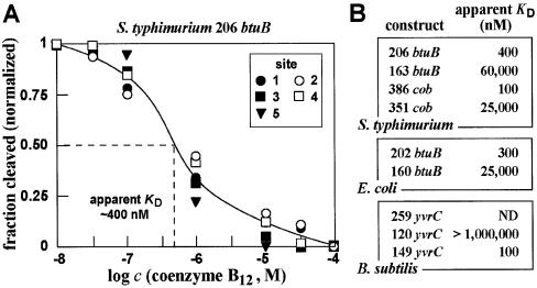
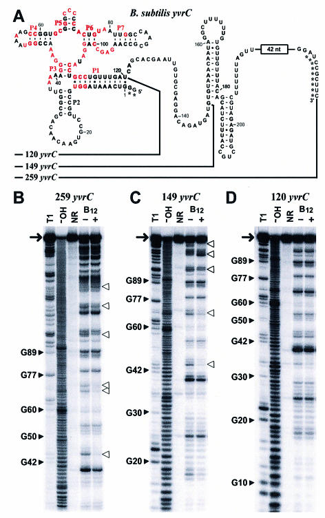
Recent studies have begun to reveal that numerous fundamental metabolic pathways in bacteria are regulated by riboswitches residing within certain messenger RNAs. These riboswitches selectively bind metabolites and modulate gene expression in response to changing ligand concentrations. Previously, we provided evidence that the btuB mRNAs of Escherichia coli and Salmonella typhimurium each carry a coenzyme B12-dependent riboswitch that causes repressed translation of the encoded cobalamin-transport protein at elevated coenzyme concentrations. Herein, we use a phylogenetic analysis to define a consensus sequence and secondary structure model for the ligand- binding domain of this riboswitch class. RNA structures that conform to this model are widespread in both Gram-positive and Gram-negative organisms. In addition, we find that the 5'-untranslated region (5'-UTR) of the cobalamin biosynthesis (cob) operon of S.typhimurium carries an RNA motif that matches this consensus sequence. Biochemical and genetic characterization of this motif confirms that the RNA directly binds coenzyme B12, and that it likely serves as a genetic control element for regulating expression of the 25-gene operon for cobalamin production in this pathogen.


Figures

Figure 1
Figure 1
Sequence alignment of putative coenzyme B12 riboswitches. Conserved sequence and base pairing elements are highlighted for 43 of 92 representative motifs identified by searching GenBank (see Materials and methods). The domain’s strand orientation (minus strand unless otherwise noted) and nucleotide number are provided (Position) for the 5′ nucleotide of the first interior UG base pair of stem P1 (asterisk). Conserved base-pairing interactions are colored, and are labeled via the secondary structure diagram at the top. Numbers in the alignment represent stretches of nucleotides that are not depicted. The region between P7 and P10 is highly variable, but always contains a putative P8 pairing. Only one example of a riboswitch-like domain is shown for each organism. For organisms with multiple riboswitches, the total number is indicated (#). A full alignment of all sequences is provided as supplementary information and has been submitted to the Rfam database (16). The Sorghum and Leishmania entries (&) are genomic clones from unfinished eukaryotic genomes. As such, these riboswitch sequences might be derived from bacterial contamination. The top four sequences are examined in this study.
Figure 2
Figure 2
Consensus sequence and secondary-structure model for the metabolite-binding domain of the B12 class of riboswitches. (A) Red nucleotides depict bases whose identities are conserved in at least 90% of the representatives of the phylogeny assembled by database searching (see Fig. 1). (B) Sequence and revised secondary-structure model for the B12 riboswitch from the 5′-UTR of the E.coli btuB gene. In-line structural probing data were taken from a study published previously (10). Although the portion of the RNA corresponding to the most highly conserved domain throughout evolution ends at nucleotide 160, the sequences through nucleotide 202 are required for maximal binding affinity (see Fig. 4). The previously identified B12 box (12,14) resides between nucleotides 140 and 160.
Figure 3
Figure 3
Structure and function of two B12 riboswitch domains from S.typhimurium. Sequence and secondary structure models for the B12 aptamer domains residing in the 5′-UTRs of the btuB mRNA (A) and the cob operon mRNA (B) from S.typhimurium. Constructs are named according to the last nucleotide derived from the respective UTR. Asterisks identify nucleotides that were added to facilitate transcription in vitro. Nucleotides denoted in red correspond to the consensus sequences as depicted in Figure 2A. Corresponding in-line probing analyses for the btuB (C) and cob (D) mRNA leader sequences are presented. T1, OH and NR identify RNAs that were partially digested with RNase T1 (cleavage after G residues), those incubated at elevated pH, or those that were not reacted, respectively. RNAs in the remaining lanes were incubated for ∼40 h in the absence of ligand (–), or in the presence of 0.1 or 1 mM coenzyme B12. Filled and open arrowheads identify selected RNase T1 digestion products, and sites of coenzyme B12-mediated structure modulation, respectively. The numbered sites of modulation for the btuB sequence were used subsequently for analysis of apparent KD (Fig. 4A).
Figure 4
Figure 4
Measurements of binding affinities of various coenzyme B12 aptamers. (A) Representative plot of the ligand concentration-dependent modulation of spontaneous RNA cleavage using the 206 btuB fragment from S.typhimurium. The fraction of RNA cleaved at sites 1–5 (Fig. 3C) was normalized against the fraction cleaved at each site when incubated in the absence of coenzyme B12. Site 5 is not present in the preceding image, but is present in the larger RNA construct used for this KD analysis (data not shown). (B) Comparison of the apparent KD values for various coenzyme B12 aptamer constructs from several bacteria. Values were derived for each construct using the approach described in (A).
Figure 5
Figure 5
A variant riboswitch aptamer retains selective binding of coenzyme B12. (A) The B12 riboswitch-like domain residing upstream of the yvrC gene of B.subtilis. The 149 yvrC and 120 yvrC constructs carry an additional two nucleotides preceding position 1 to facilitate efficient transcription in vitro. Similarly, the 259 yvrC construct carried an additional sequence (GGAAAAACGGAUACGAAU) residing immediately upstream from nucleotide 1, wherein the first G residue was altered from the natural nucleotide identity. Asterisks identify non-natural nucleotides. (BD) In-line probing analyses of three RNA fragments of the putative yvrC 5′-UTR of B.subtilis. Details are as described in the legend to Figure 3.

References

    1. Winkler W.C. and Breaker,R.R. (2003) Genetic control by metabolite-binding riboswitches. Chembiochem, 4, 1024–1032. - PubMed
    1. Winkler W.C. Cohen-Chalamish,S. and Breaker,R.R. (2002) An mRNA structure that controls gene expression by binding FMN. Proc. Natl Acad. Sci. USA, 99, 15908–15913. - PMC - PubMed
    1. Mironov A.S., Gusarov,I., Rafikov,R., Lopez,L.E., Shatalin,K., Kreneva,R.A., Perumov,D.A. and Nudler,E. (2002) Sensing small molecules by nascent RNA: a mechanism to control transcription in bacteria. Cell, 111, 747–756. - PubMed
    1. Winkler W., Nahvi,A. and Breaker,R.R. (2002) Thiamine derivatives bind messenger RNAs directly to regulate bacterial gene expression. Nature, 419, 952–956. - PubMed
    1. Mandal M., Boese,B., Barrick,J.E., Winkler,W.C. and Breaker,R.R. (2003) Riboswitches control fundamental biochemical pathways in Bacillus subtilis and other bacteria. Cell, 113, 577–586. - PubMed

Publication types

MeSH terms