

doi: 10.1126/science.1088493.
Crystal structure of biotin synthase, an S-adenosylmethionine-dependent radical enzyme
Affiliations
- PMID: 14704425
- PMCID: PMC1456065
- DOI: 10.1126/science.1088493
Item in Clipboard
Crystal structure of biotin synthase, an S-adenosylmethionine-dependent radical enzyme
Science.
.
Abstract
The crystal structure of biotin synthase from Escherichia coli in complex with S-adenosyl-L-methionine and dethiobiotin has been determined to 3.4 angstrom resolution. This structure addresses how "AdoMet radical" or "radical SAM" enzymes use Fe4S4 clusters and S-adenosyl-L-methionine to generate organic radicals. Biotin synthase catalyzes the radical-mediated insertion of sulfur into dethiobiotin to form biotin. The structure places the substrates between the Fe4S4 cluster, essential for radical generation, and the Fe2S2 cluster, postulated to be the source of sulfur, with both clusters in unprecedented coordination environments.
Figures
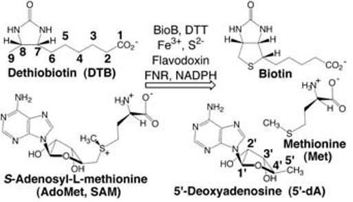
Overall reaction catalyzed by BioB.
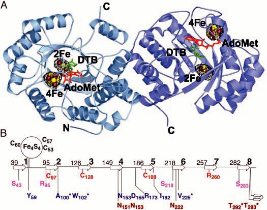
(A) Structure of BioB, FeS clusters, and bound AdoMet and DTB. BioB exists as a homodimer in solution (11) and we find two possible dimeric relationships between BioB monomers in the crystal. The dimer shown here buries 17.6% of the monomer surface area (13,249 Å2) and is likely to be physiologically relevant. An anomalous Fourier electron density map, calculated with data collected at the Fe absorption peak wavelength (1.73827 Å) and phases from the polypeptide portion of the model, is contoured at 3σ in black mesh. These electron density peaks represent the positions of the four FeS clusters in this dimeric structure. There are no other features of a similar size in the electron density map. The FeS clusters are shown as ball-and-stick representations, with brown Fe atoms and yellow S atoms. In addition, we find one AdoMet (red) and one DTB (green) per subunit. Figures 1A and 2 were prepared with PyMOL (42). (B) Topology diagram of the BioB TIM barrel showing the location of important residues with respect to the β strands (arrows, numbered 1 to 8). The numbers to the left of each β strand correspond to the N-terminal residue of that secondary structure element. Ligands to the Fe4S4 cluster are in black, ligands to the Fe2S2 cluster are in red, and residues that contact the Fe2S2 cluster ligand Arg260 are in pink. AdoMet contacts (blue) include Ala100, Trp102, and Arg173, which form hydrogen bonds to the amino acid moiety; Asp155 and Asn153, which form hydrogen bonds to the ribose hydroxyl groups (Fig. 2A); Tyr59 and Ile192, which stack against the adenine ring (Fig. 2A); and Val225, which forms backbone hydrogen bonds to the adenine ring. Residues in position to form hydrogen bonds to DTB (brown) include Asn151, Asn153, and Asn222, which contact the DTB ureido ring (Fig. 2C), and Thr292 and Thr293, which contact the carboxylate tail. Asterisks denote main-chain interactions.
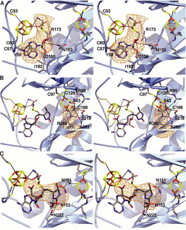
(A) Stereo view of the Fe4S4 cluster with AdoMet bound. Conserved side-chain contacts between BioB and AdoMet are indicated, and AdoMet is shown in a simulated annealing omit map contoured at 4.5σ (orange). DTB is omitted for clarity. Color code: C, gray; O, red; N, blue; S, yellow; Fe, brown. (B) Stereo view of the active site, focusing on the Fe2S2 cluster and its ligands. The unusual Arg260 ligand is shown in a simulated annealing omit map contoured at 4.5σ. In addition to the Fe2S2 cluster, Arg260 interacts with Ser43, Ser218, Ser283, and Arg95. Also shown are the positions of the Fe4S4 cluster, AdoMet, and DTB with respect to the Fe2S2 cluster. (C) Stereo view of DTB interacting with AdoMet and conserved residues Asn222, Asn151, and Asn153 in the active site. Potential hydrogen bonds between Asn222 and DTB are drawn as dashed lines. The stacking of the carboxylate tail of DTB and the adenine ring of AdoMet is visible in the orientation, although contacts with Thr292 and Thr293 are not. DTB is shown in a simulated annealing omit map contoured at 4.0σ.
References
Publication types
MeSH terms
Substances
Associated data
- Actions
Grants and funding
LinkOut - more resources
Full Text Sources
Other Literature Sources
Molecular Biology Databases
