. 2004 Jan 30;303(5658):672-6.
doi: 10.1126/science.1093686. Epub 2004 Jan 2.

RNAi-mediated targeting of heterochromatin by the RITS complex

Affiliations

RNAi-mediated targeting of heterochromatin by the RITS complex

André Verdel et al. Science. .

Erratum in

  • Science. 2013 Oct 11;342(6155):193

Abstract

RNA interference (RNAi) is a widespread silencing mechanism that acts at both the posttranscriptional and transcriptional levels. Here, we describe the purification of an RNAi effector complex termed RITS (RNA-induced initiation of transcriptional gene silencing) that is required for heterochromatin assembly in fission yeast. The RITS complex contains Ago1 (the fission yeast Argonaute homolog), Chp1 (a heterochromatin-associated chromodomain protein), and Tas3 (a novel protein). In addition, the complex contains small RNAs that require the Dicer ribonuclease for their production. These small RNAs are homologous to centromeric repeats and are required for the localization of RITS to heterochromatic domains. The results suggest a mechanism for the role of the RNAi machinery and small RNAs in targeting of heterochromatin complexes and epigenetic gene silencing at specific chromosomal loci.


Figures

Fig. 1
Fig. 1
Purification of Chp1-TAP and identification of associated proteins. Extracts from a Chp1-TAP strain and an untagged control strain were purified by the TAP procedure and applied to a 4 to 12% polyacrylamide gel, which was stained with colloidal Coomassie blue (A). The bands in the Chp1-TAP purification were excised from the gel and sequenced by tandem mass spectrometry (22). The identity of each band is based on multiple sequenced peptides and is indicated on the right. *Residual GST-TEV, the protease used for elution from the first affinity column. (B) The Chp1-TAP protein was fully functional for silencing of a centromeric imr::ura4+ reporter gene as indicated by wild-type levels of growth on 5-FOA medium, which only allows growth when ura4+ is silenced. N/S, nonselective medium. (C) Schematic diagram showing the subunits of the RITS complex and their conserved motifs. The chromodomain (ChD) in Chp1, the PAZ and PIWI domains in Ago1, and a region of sequence similarity between Tas3 and the mouse OTT (ovary testis transcribed) protein are indicated.
Fig. 2
Fig. 2
Purification of the RITS complex by using a Tas3-TAP strain and the requirement of tas3+ in silencing and methylation of H3-K9 and Swi6 localization. Western blot showing that (A) the Tas3-TAP and Chp1-TAP proteins are expressed to similar levels and (B) growth assays showing that Tas3-TAP displays wild-type levels of silencing for a centromeric imrIR::ura4+ reporter gene. (C) Tas3-TAP was purified, and silver-stained protein bands were sequenced by tandem mass spectrometry. *GST-TEV. (D) In tas3Δ cells, silencing of a ura4+ reporter gene inserted at the centromeric repeats (imr1R::ura4+ and otr1R::ura4+) is lost, but silencing of the same reporter gene at the silent mating-type interval (Kint2::ura4+) is unaffected. Loss of silencing in sir2Δ, chp1Δ, and ago1Δ is shown for comparison. Loss of silencing results in loss of growth on counterselective 5-FOA medium. (E) ChIP experiments showing that in tas3Δ cells methylation of histone H3-K9 and localization of Swi6 to a ura4+ reporter gene inserted at otr1R and imr1R centromeric repeats is abolished. In contrast, deletion of tas3+ has little or no effect on H3-K9 methylation and Swi6 localization (Kin2::ura4+). ChIP analysis and quantification were performed as described previously (26). The ratios of ura4+ or cen signals to ura4DS/E-minigene signal present in the immunoprecipitated DNA (ChIP) and whole-cell extracts (WCE) were used to calculate fold enrichment shown underneath each lane.
Fig. 3
Fig. 3
Dicer-dependent association of RITS with siRNAs. (A) Small RNAs of ~22 to 25 nt copurify with Chp1-TAP. RNAs isolated from untagged control (−) and Chp1-TAP (+) strains were 3′ end-labeled with [5′-32P]pCp and separated on 15% denaturing urea polyacrylamide gel. Lane 1, [γ-32P]ATP–labeled RNA markers (Ambion); lanes 2 and 3, labeling of RNA from whole-cell extract (WCE) (~1/2500 of input); lanes 3 and 4, labeling of RNAs after purification. Bracket on the right side indicates the position of small RNAs specifically associated with Chp1-TAP. (B) Copurification of small RNAs with Tas3-TAP. (C) No small RNAs are associated with RITS purified from dcr1Δ cells. Parallel purifications were performed from an untagged (control, lane 1) strain as well as chp1-TAP, dcr1+ (lane 2) and chp1-TAP, dcr1Δ (lane 3) cells, and the associated RNAs were [5′-32P]pCp labeled (compare lanes 2 and 3, bracket). (D) Northern blot showing that siRNAs associated with RITS hybridize to 32P-labeled probes corresponding to centromeric repeat sequences. RNA from untagged control (lane1) and Chp1-TAP cells (lane 2), purified as described in (B), was separated on a denaturing gel and electrotransferred to a nylon membrane (22). DNA oligonucleotides with sequence complementary to the 12 heterochromatic siRNAs identified by Reinhart and Bartel (16) were 5′ labeled with [γ-32P]ATP and used as probes for the Northern blot. (E) Southern blot showing that RITS contains siRNAs complementary to the outer centromeric repeats (otr). dg (lanes 2 and 4) and dh (lane 3) repeats, actin (lane 5), and LTRs (lane 6) were amplified by polymerase chain reaction (PCR) from genomic DNA, separated on 1.1% agarose gel, and transferred to nylon membrane. 32P-labeled RITS siRNAs, obtained by labeling RNAs as described in (A), were separated on a denaturing urea gel, eluted, and used as probes for the blot.
Fig. 4
Fig. 4
The RNAi pathway is required for localization of RITS to heterochromatin. (A) ChIP experiments showing that Tas3-TAP is localized to centromeric heterochromatin in an RNAi-dependent manner. Tas3-TAP is associated with ura4+ inserted at the otr centromeric repeats (otr1::ura4+, left panels) and with native centromeric repeat sequences (cen, right panels) in wild-type (wt) but not ago1Δ, dcr1Δ, or rdp1Δ cells. The ura4DS/E-minigene at the endogenous euchromatic location is used as a control. (B) The RNAi pathway is required for the localization of Chp1-(Flag)3 to centromeric heterochromatin. (C) Tas3 is required for the localization of Chp1-(Flag)3 to heterochromatin. Immunoprecipitations were carried out using a Flag-specific antibody from tas3+ and tas3Δ cells. (D) Tas3 is associated with ura4+ inserted at the imr centromeric region (imr1::ura4+). WCE, whole-cell extract. Fold enrichment values are shown underneath each lane.
Fig. 5
Fig. 5
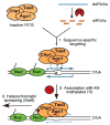
A model for siRNA-dependent initiation of heterochromatin assembly by RITS. The RITS complex is programmed by Dcr1-produced siR-NAs to target specific chromosome regions by sequence-specific interactions involving either siRNA-DNA or siRNA-nascent transcript (blue arrows) base pairing. Nuc, nucleosome; red triangle, K9-methylation on the amino terminus of histone H3. See text for further discussion and references.

Similar articles

Cited by

References

    1. Grewal SIS. J Cell Physiol. 2000;184:311. - PubMed
    1. Grewal SIS, Moazed D. Science. 2003;301:798. - PubMed
    1. Partridge JF, Scott KS, Bannister AJ, Kouzarides T, Allshire RC. Curr Biol. 2002;12:1652. - PubMed
    1. Zilberman D, Cao X, Jacobsen SE. Science. 2003;299:716. - PubMed
    1. Matzke M, Matzke AJM, Kooter JM. Science. 2001;293:1080. - PubMed

Publication types

MeSH terms