. 2004 Jan 5;199(1):35-46.
doi: 10.1084/jem.20030204.

Granulocyte CEACAM3 is a phagocytic receptor of the innate immune system that mediates recognition and elimination of human-specific pathogens

Affiliations

Granulocyte CEACAM3 is a phagocytic receptor of the innate immune system that mediates recognition and elimination of human-specific pathogens

Tim Schmitter et al. J Exp Med. .

Abstract

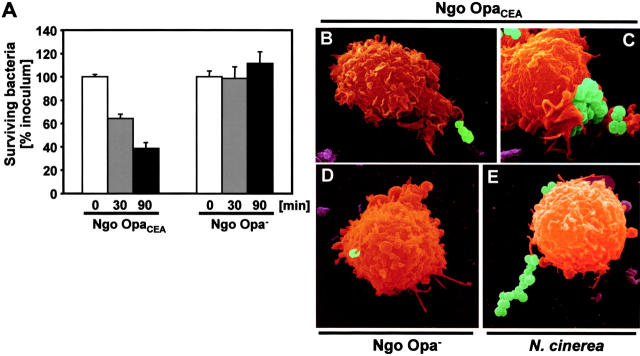
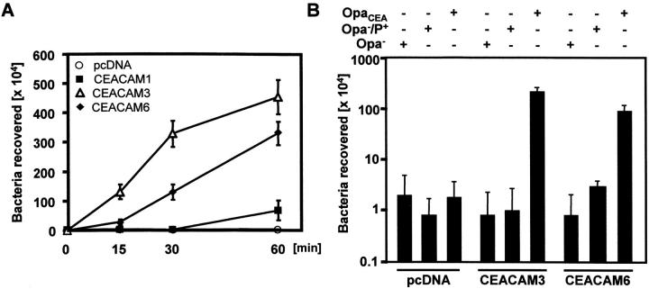
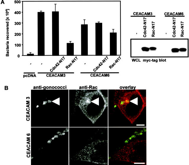
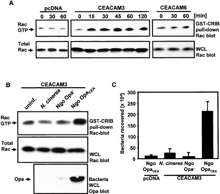
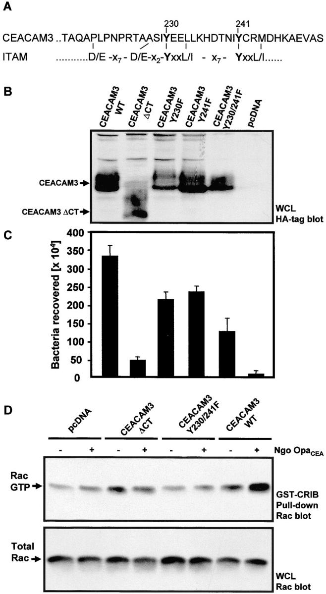
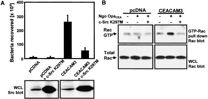
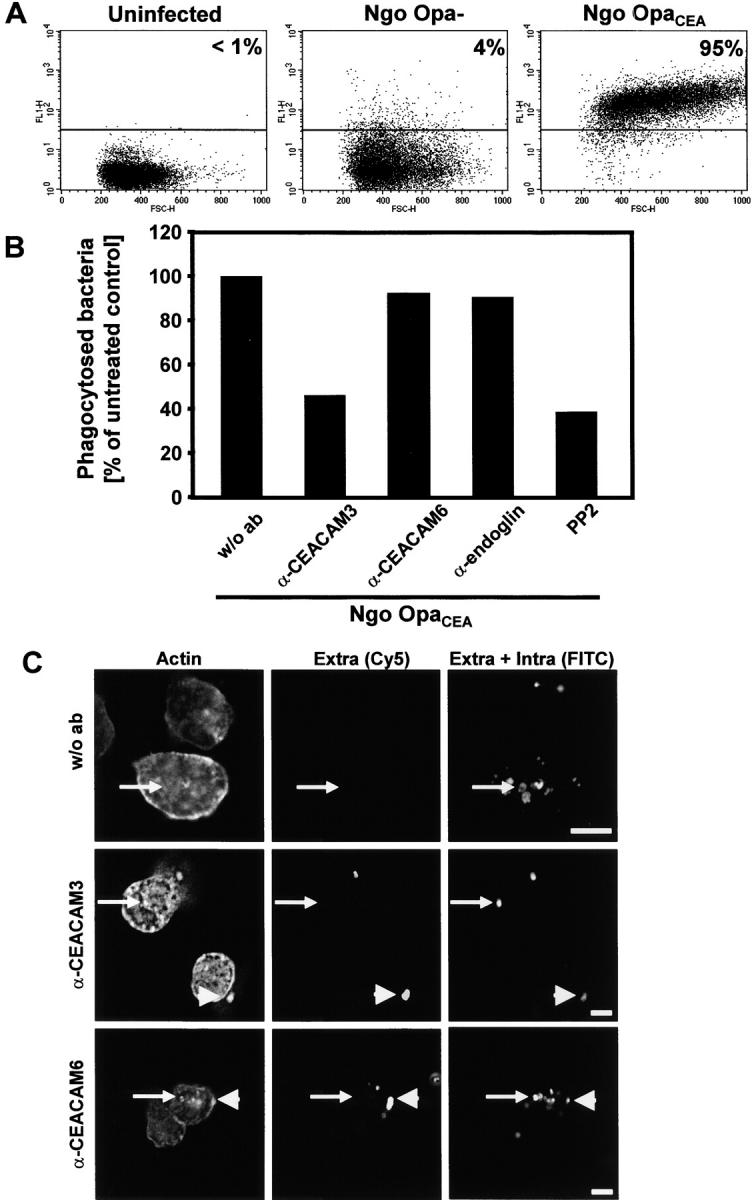
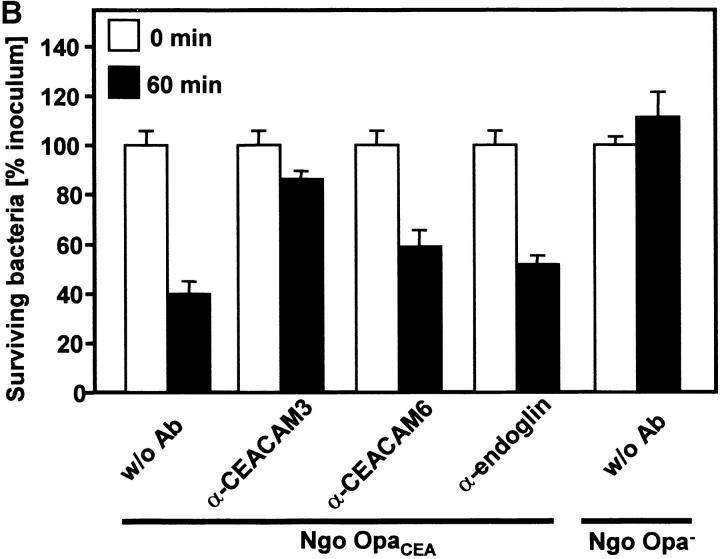
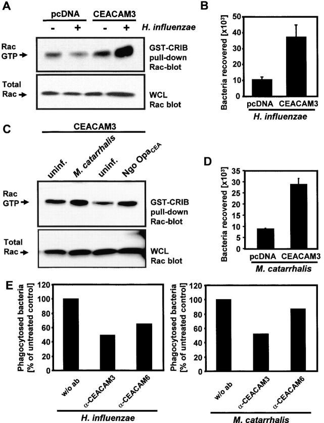
Carcinoembryonic antigen-related cell adhesion molecules (CEACAMs) are used by several human pathogens to anchor themselves to or invade host cells. Interestingly, human granulocytes express a specific isoform, CEACAM3, that participates together with CEACAM1 and CEACAM6 in the recognition of CEACAM-binding microorganisms. Here we show that CEACAM3 can direct efficient, opsonin-independent phagocytosis of CEACAM-binding Neisseria, Moraxella, and Haemophilus species. CEACAM3- but not CEACAM6-mediated uptake is blocked by dominant-negative versions of the small GTPase Rac. Moreover, CEACAM3 engagement triggers membrane recruitment and increased GTP loading of Rac that are not observed upon bacterial binding to CEACAM6. Internalization and Rac stimulation are also inhibited by compromising the integrity of an immunoreceptor tyrosine-based activation motif (ITAM)-like sequence in the cytoplasmic tail of CEACAM3 or by interference with Src family protein tyrosine kinases that phosphorylate CEACAM3. In contrast to interfering with CEACAM6, blockage of CEACAM3-mediated events reduces the ability of primary human granulocytes to internalize and eliminate CEACAM-binding bacteria, indicating an important role of CEACAM3 in the control of human-specific pathogens by the innate immune system.


Figures

Figure 1.
Figure 1.
OpaCEA-expressing N. gonorrhoeae are efficiently recognized and eliminated by human granulocytes via lamellipodia-like membrane protrusions. (A) Granulocytes were incubated with OpaCEA gonococci (Ngo OpaCEA) or nonopaque, piliated gonococci (Ngo Opa), respectively. At the indicated times, the number of viable gonococci was determined and expressed as the percentage of the initial inoculum. The graph shows mean values ± SDs of a representative experiment done in triplicate. Equivalent results were obtained with granulocytes isolated from four different donors. (B–E) Pseudocolored scanning electron micrographs of granulocytes infected for 1 h with OpaCEA gonococci (B and C), nonopaque N. gonorrhoeae (D), or nonpathogenic N. cinerea (E).
Figure 2.
Figure 2.
Transient expression of CEACAMs in 293 cells allows interaction with OpaCEA gonococci. (A) 293 cells were transfected with CEACAM1, CEACAM3, CEACAM6, or the empty expression vector (pcDNA). After 2 d, cells were infected with OpaCEA gonococci. At the indicated time points, the number of internalized bacteria was determined by gentamicin protection assays. The graph shows mean values ± SDs of three independent experiments done in triplicate. (B) Transfected 293 cells were infected with nonopaque (Opa), nonopaque, piliated (Opa/P+), or OpaCEA-expressing (OpaCEA) gonococci for 1 h and analyzed in gentamicin protection assays. The graph shows mean values ± SDs of two independent experiments done in triplicate.
Figure 3.
Figure 3.
CEACAM3- but not CEACAM6-mediated internalization of gonococci depends on the small GTPase Rac. (A) 293 cells cotransfected with Cdc42 N17 or Rac N17 and either CEACAM3 or CEACAM6 were infected with OpaCEA gonococci, and the number of internalized bacteria was determined in gentamicin protection assays. The graph shows mean values ± SDs of three independent experiments done in triplicate. Western blotting of whole cell lysates (WCL) with anti–myc tag antibody demonstrates expression of transfected Cdc42 N17 and Rac N17, respectively. (B) 293 cells, transfected with CEACAM3 or CEACAM6, were infected with OpaCEA N. gonorrhoeae for 1 h, fixed, and stained with polyclonal antibodies against gonococci and monoclonal antibodies against Rac. In CEACAM3-transfected cells, Rac is recruited to the site of bacteria–cell interaction (arrowhead).
Figure 4.
Figure 4.
CEACAM3 engagement by OpaCEA gonococci results in Rac stimulation. (A) 293 cells transfected with the indicated constructs were infected with OpaCEA N. gonorrhoeae for different time points. GTP-bound Rac was isolated using the Rac-binding domain of PAK (GST-CRIB) and detected by Western blotting with anti-Rac antibodies (top). Whole cell lysates (WCL) were also probed with anti-Rac antibodies (bottom). (B) CEACAM3-expressing 293 cells were left uninfected or were infected with N. cinerea, piliated, nonopaque N. gonorrhoeae (Ngo Opa) or OpaCEA N. gonorrhoeae (Ngo OpaCEA). GST-CRIB pull-downs (top) or whole cell lysates (middle) were probed with anti-Rac antibodies. Lysates of the bacteria were probed with monoclonal anti-Opa antibody (bottom). (C) 293 cells transfected with the empty control vector (pcDNA) or CEACAM3 were infected with the bacteria employed in B and analyzed in gentamicin protection assays. The graph shows mean values ± SDs of three independent experiments done in triplicate.
Figure 5.
Figure 5.
The ITAM-like sequence of CEACAM3 is critical for bacterial internalization and Rac stimulation. (A) Primary sequence of the COOH-terminal cytoplasmic domain of CEACAM3 containing the ITAM-like sequence. The ITAM consensus (31) is shown below with the critical tyrosine residues in bold. (B) 293 cells were transfected with the control vector (pcDNA) or the indicated HA-tagged CEACAM3 constructs. Whole cell lysates (WCL) were analyzed with anti–HA tag antibody. (C) Transfected cells as in B were infected with OpaCEA-expressing gonococci for 60 min and analyzed in gentamicin protection assays. The graph shows mean values ± SDs of three to four independent experiments done in triplicate. (D) Cells were transfected as in B, and samples were lysed before or after 60 min of infection with OpaCEA N. gonorrhoeae. GST-CRIB pull-down assays (top) or whole cell lysates (WCL; bottom) of the samples were analyzed with monoclonal anti-Rac antibody.
Figure 6.
Figure 6.
Src kinases connect CEACAM3 engagement with Rac stimulation. (A) 293 cells were transfected with the control vector (pcDNA), CEACAM3, or cotransfected with a kinase-inactive mutant of c-Src (c-Src K297M), infected with OpaCEA gonococci, and employed in gentamicin protection assays. The graph shows mean values ± SDs of two independent experiments done in triplicate. Whole cell lysates (WCL) of the samples were analyzed for Src expression (bottom). (B) Lysates of cells as in A were prepared before and after infection with OpaCEA gonococci. GST-CRIB pull-down assays (top) or whole cell lysates (WCL; bottom) of the samples were analyzed with monoclonal anti-Rac antibody.
Figure 7.
Figure 7.
CEACAM3 is critical for uptake of CEACAM-binding bacteria by primary human granulocytes. (A) Granulocytes were incubated with FITC-labeled OpaCEA gonococci (Ngo OpaCEA) or nonopaque, piliated gonococci (Ngo Opa), respectively, or left uninfected. After 20 min, phagocytosis was measured by FACS® analysis. Values represent the percentage of granulocytes containing FITC-labeled bacteria from a representative experiment. Similar values were obtained with granulocytes from three different donors. (B) Phagocytosis of OpaCEA bacteria was analyzed as above in the absence or presence of 20 μg of the indicated monoclonal antibodies or 10 μM of the Src PTK inhibitor PP2. The graph shows mean values of a representative experiment done in triplicate. Equivalent results were obtained with granulocytes isolated from three different donors. (C) Human granulocytes were infected with FITC-labeled OpaCEA gonococci in the presence or absence of 20 μg of the indicated monoclonal antibodies for 60 min. Cells were differentially stained for extracellular (arrowheads) and intracellular bacteria (small arrow). Cellular actin was visualized with phalloidin-TRITC.
Figure 8.
Figure 8.
CEACAM3 and Rac stimulation are required for efficient uptake and elimination of OpaCEA gonococci by primary human granulocytes. (A) Granulocytes were either untreated or pretreated with the indicated amounts of TAT-LacZ, TAT-RacN17, or TAT-Cdc42N17 and infected with FITC-labeled OpaCEA gonococci (Ngo OpaCEA). Phagocytosis was determined by FACS® analysis, and the graphs show the result of a representative experiment. Similar results were obtained with granulocytes isolated from three different donors. (B) Granulocytes were incubated with OpaCEA gonococci (Ngo OpaCEA) or nonopaque, piliated gonococci (Ngo Opa), respectively, in the absence or presence of 20 μg of the indicated antibodies. Before and after 60 min of incubation, aliquots of the samples were plated on GC agar to determine the number of viable gonococci. The graph shows mean values ± SDs of a representative experiment done in triplicate. Equivalent results were obtained with granulocytes isolated from three different donors.
Figure 8.
Figure 8.
CEACAM3 and Rac stimulation are required for efficient uptake and elimination of OpaCEA gonococci by primary human granulocytes. (A) Granulocytes were either untreated or pretreated with the indicated amounts of TAT-LacZ, TAT-RacN17, or TAT-Cdc42N17 and infected with FITC-labeled OpaCEA gonococci (Ngo OpaCEA). Phagocytosis was determined by FACS® analysis, and the graphs show the result of a representative experiment. Similar results were obtained with granulocytes isolated from three different donors. (B) Granulocytes were incubated with OpaCEA gonococci (Ngo OpaCEA) or nonopaque, piliated gonococci (Ngo Opa), respectively, in the absence or presence of 20 μg of the indicated antibodies. Before and after 60 min of incubation, aliquots of the samples were plated on GC agar to determine the number of viable gonococci. The graph shows mean values ± SDs of a representative experiment done in triplicate. Equivalent results were obtained with granulocytes isolated from three different donors.
Figure 9.
Figure 9.
M. catarrhalis and H. influenzae are recognized and phagocytosed via CEACAM3. (A) Empty control vector (pcDNA) or CEACAM3-transfected 293 cells were left uninfected or were infected with unencapsulated H. influenzae strain RD for 60 min. GST-CRIB pull-downs (top) or whole cell lysates (bottom) were probed with anti-Rac antibodies. (B) Cells as in A were infected with H. influenzae strain RD and analyzed in gentamicin protection assays. The graph shows mean values ± SDs of two independent experiments done in triplicate. (C) CEACAM3-transfected 293 cells were left uninfected or were infected with M. catarrhalis strain 11994 or with OpaCEA gonococci (Ngo OpaCEA) for 60 min. GST-CRIB pull-downs (top) or whole cell lysates (bottom) were probed with anti-Rac antibodies. (D) 293 cells transfected with the empty control vector (pcDNA) or CEACAM3 were infected with M. catarrhalis for 60 min and analyzed in gentamicin protection assays. The graph shows mean values ± SDs of two independent experiments done in triplicate. (E) Human granulocytes were infected with FITC-labeled M. catarrhalis or H. influenzae, respectively, in the presence or absence of the indicated antibodies for 20 min and analyzed by FACS® for intracellular bacteria. The graphs show the results of representative experiments. Similar results were obtained with granulocytes isolated from two different donors.

References

    1. Ryan, K.J. 1990. Neisseria including Branhamella. Medical Microbiology—An Introduction to Infectious Diseases. J.C. Sheris, editor. Elsevier Science Publishing Co. Inc., New York/Amsterdam/ London. 343–356.
    1. Hedges, S.R., M.S. Mayo, J. Mestecky, E.W. Hook, III, and M.W. Russell. 1999. Limited local and systemic antibody responses to Neisseria gonorrhoeae during uncomplicated genital infections. Infect. Immun. 67:3937–3946. - PMC - PubMed
    1. Schmidt, K.A., H. Schneider, J.A. Lindstrom, J.W. Boslego, R.A. Warren, L. Van de Verg, C.D. Deal, J.B. McClain, and J.M. Griffiss. 2001. Experimental gonococcal urethritis and reinfection with homologous gonococci in male volunteers. Sex. Transm. Dis. 28:555–564. - PubMed
    1. Virji, M., K. Makepeace, D.J.P. Ferguson, and S.M. Watt. 1996. Carcinoembryonic antigens (CD66) on epithelial cells and neutrophils are receptors for Opa proteins of pathogenic Neisseriae. Mol. Microbiol. 22:941–950. - PubMed
    1. Gray-Owen, S.D., C. Dehio, A. Haude, F. Grunert, and T.F. Meyer. 1997. CD66 carcinoembryonic antigens mediate interactions between Opa-expressing Neisseria gonorrhoeae and human polymorphonuclear phagocytes. EMBO J. 16:3435–3445. - PMC - PubMed

Publication types