Comparative Study
. 2004 Jan;14(1):1-10.
doi: 10.1101/gr.1717804.

Developmental roles of pufferfish Hox clusters and genome evolution in ray-fin fish

Affiliations
Comparative Study

Developmental roles of pufferfish Hox clusters and genome evolution in ray-fin fish

Angel Amores et al. Genome Res. 2004 Jan.

Abstract

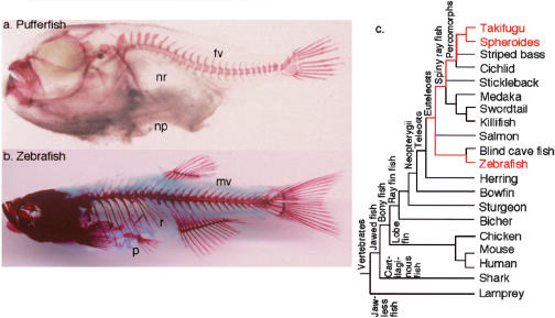
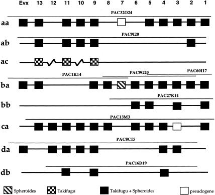
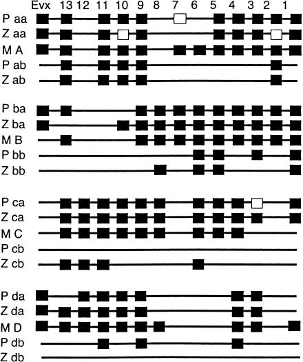
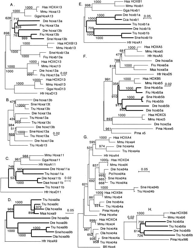
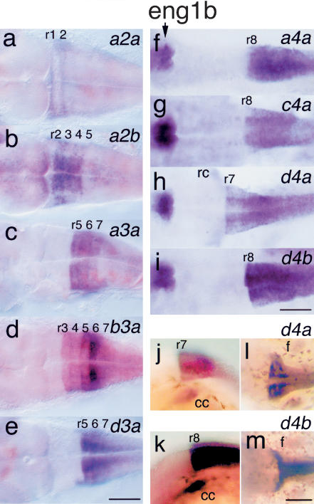
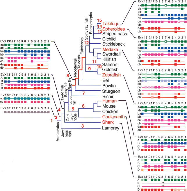
The pufferfish skeleton lacks ribs and pelvic fins, and has fused bones in the cranium and jaw. It has been hypothesized that this secondarily simplified pufferfish morphology is due to reduced complexity of the pufferfish Hox complexes. To test this hypothesis, we determined the genomic structure of Hox clusters in the Southern pufferfish Spheroides nephelus and interrogated genomic databases for the Japanese pufferfish Takifugu rubripes (fugu). Both species have at least seven Hox clusters, including two copies of Hoxb and Hoxd clusters, a single Hoxc cluster, and at least two Hoxa clusters, with a portion of a third Hoxa cluster in fugu. Results support genome duplication before divergence of zebrafish and pufferfish lineages, followed by loss of a Hoxc cluster in the pufferfish lineage and loss of a Hoxd cluster in the zebrafish lineage. Comparative analysis shows that duplicate genes continued to be lost for hundreds of millions of years, contrary to predictions for the permanent preservation of gene duplicates. Gene expression analysis in fugu embryos by in situ hybridization revealed evolutionary change in gene expression as predicted by the duplication-degeneration-complementation model. These experiments rule out the hypothesis that the simplified pufferfish body plan is due to reduction in Hox cluster complexity, and support the notion that genome duplication contributed to the radiation of teleosts into half of all vertebrate species by increasing developmental diversification of duplicate genes in daughter lineages.


Figures

Figure 1
Figure 1
Morphology and phylogeny. Pufferfish lack ribs and pelvis and have few vertebrae, as revealed by Alizarin red staining of the skeleton of the pufferfish Takifugu rubripes (a) and the zebrafish Danio rerio (b). (c) A phylogenetic tree for vertebrates (see Nelson 1994), times according to Hedges (2002), Hedges and Kumar (2002), and Santini and Tyler (1999). (fv) Few vertebrae; (mv) many vertebrae;(np) no pelvic appendage; (nr) no ribs; (p) pelvic apparatus; (r) ribs.
Figure 2
Figure 2
Genomic organization of pufferfish Hox clusters. Hox paralog group shown along the top and cluster designation at left. Plate and well numbers of PACs as well as their genomic extent are sketched. (Filled squares) Genes present in both S. nephalus and T. rubripes. (Squares with diagonal lines) Genes found in S. nephalus butnot T. rubripes. (Checkered squares) Genes found in T. rubripes butnot S. nephalus. (Empty squares) Pseudogenes.
Figure 3
Figure 3
Comparative genomics of pufferfish (P), zebrafish (Z), and mouse (M) Hox clusters. Paralog group shown along the top and cluster designation at left.
Figure 4
Figure 4
Phylogenetic relationships of pufferfish Hox clusters. Neighbor-joining trees are based on amino acid sequences as described (Amores etal. 1998). (A) Paralog group 13, exon 1, and exon 2. The tree is as expected if Hox cluster duplication occurred before the divergence of pufferfish and zebrafish lineages. (B) Paralog group 13, exon 2 only (due to limited sequence availability). The tree shows that Hoxa13c is the sister group to Hoxa13a. (C) Hoxa11a of pufferfish and zebrafish group as sisters, but Hoxa11b orthologs do not. (D) Hoxa9 tree strongly supports duplication before lineage divergence, and the close similarity of the Hoxaa and Hoxac clusters. (E,F) The Hoxb1 and paralog group-5 trees support duplication before lineage divergence. (G) The paralog group-4 tree shows rapid evolution of Hoxd4b. (H) The Hoxb6 tree supports duplication before lineage divergence. Alignments and accession numbers are available as Supplemental material. Numbers are bootstrap values per 1000 runs. (Cca) Carassius carassius, crucian carp; (Dae) Danio aequipinnatus, giantdanio; (Bfl) Branchiostoma floridae, amphioxus; (Dre) Danio rerio, zebrafish; (Gga) Gallus gallus, chicken; (Hfr) Heterodontus francisci, horned shark; (Hsa) Homo sapiens, human; (Mmu) Mus musculus mouse; (Msa) Morone saxatilis, striped bass; (Ola) Oryzias latipes, medaka; (Pma) Petromyzon marinus, lamprey; (Pol) Paralichthys olivaceus, Japanese flounder; (Sne) Spheroides nephalus, Southern pufferfish; (Tru) Takifugu rubripes, Japanese pufferfish.
Figure 5
Figure 5
Expression of Hox genes in 5-d fugu embryos. (a) Hoxa2a is expressed in an apparently novel striped pattern in r1 and r2. (b) Hoxa2b is expressed in r2-r5 as in other vertebrates. (c) Hoxa3a has an anterior boundary at the r4/r5 border as in other vertebrates. (d) Hoxb3a has strong expression in r5 and r6 and weak expression in r4 as in zebrafish, but with weak expression extending into r3 in fugu and not in zebrafish (Prince etal. 1998b). (e) Hoxd3a expression mimics that of zebrafish, with an anterior limit at the r5/r6 border, and a small lateral/ventral group of r5 cells (Prince etal. 1998b). Expression of eng1b atthe midbrain/hindbrain border serves as a marker for rhombomere position in f-i. (f) Hoxa4a is expressed with a diffuse anterior expression boundary near the r7/r8 border as in zebrafish (Prince et al. 1998b). (g) Hoxc4a is expressed with a diffuse anterior boundary, and as in zebrafish, the border lies within r7, atleastmedially, and interneurons express eng1b as in zebrafish (Force et al. 1999). (h) Hoxd4a transcript tissue distribution as in other vertebrates. (i) Hoxd4b has a more caudal anterior expression border than Hoxd4 in other vertebrates. (j) Hoxd4a in lateral view. (k) Hoxd4b in lateral view. (l) Hoxd4a in dorsal view showing fin buds. (m) Hoxd4b in dorsal view showing fin buds. Scale bar, 100 μ. (cc) Cranial crest; (f) fin buds.
Figure 6
Figure 6
The evolution of vertebrate Hox cluster genomic organization. See text for explanation.

References

    1. Ahn, S. and Tanksley, S.D. 1993. Comparative linkage maps of the rice and maize genomes. Proc. Natl. Acad. Sci. 90: 7980-7984. - PMC - PubMed
    1. Allendorf, F. and Thorgaard, G. 1984. Tetraploidy and the evolution of salmonid fishes. In Evolutionary genetics of fishes (ed. B.J. Turner), pp. 1-46. Plenum Press, New York.
    1. Allendorf, F.W., Utter, F.M., and May, B.P. 1975. Gene duplication within the family Salmonidae: II. Detection and determination of the genetic control of duplicate loci through inheritance studies and the examination of populations. In Isozymes (ed. C.L. Markert), pp. 415-432. Academic Press, New York.
    1. Amemiya, C., Amores, A., Ota, G., Mueller, D., Garraty, J., Postlethwait, J., and Litman, G. 2001. Generation of a P1 artificial chromosome library of the Southern pufferfish. Gene 272: 283-289. - PubMed
    1. Amores, A., Force, A., Yan, Y.-L., Joly, L., Amemiya, C., Fritz, A., Ho, R.K., Langel, J., Prince, V., Wang, Y.-L., et al. 1998. Zebrafish hox clusters and vertebrate genome evolution. Science 282: 1711-1714. - PubMed

WEB SITE REFERENCES

    1. http://www.ncbi.nlm.nih.gov/genome/guide/human/Q; Human Genome Resources.
    1. http://zfin.org/; The Zebrafish Information Network.
    1. http://fugu.hgmp.mrc.ac.uk/blast/; Fugu BLAST server at the UK Human Genome Mapping Project Resource Centre.
    1. http://genome.jgi-psf.org/fugu6/fugu6.home.html; Fugu rubripes at the Joint Genome Institute.

Publication types

LinkOut - more resources