. 2004 Jan 5;164(1):69-78.
doi: 10.1083/jcb.200303037.

Role of the hydrophobic domain in targeting caveolin-1 to lipid droplets

Affiliations

Role of the hydrophobic domain in targeting caveolin-1 to lipid droplets

Anne G Ostermeyer et al. J Cell Biol. .

Abstract

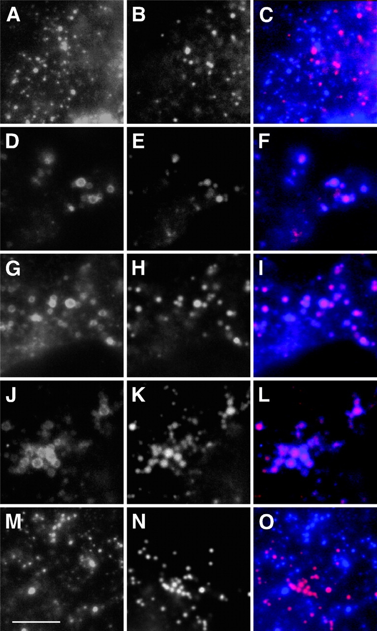
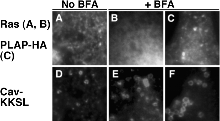
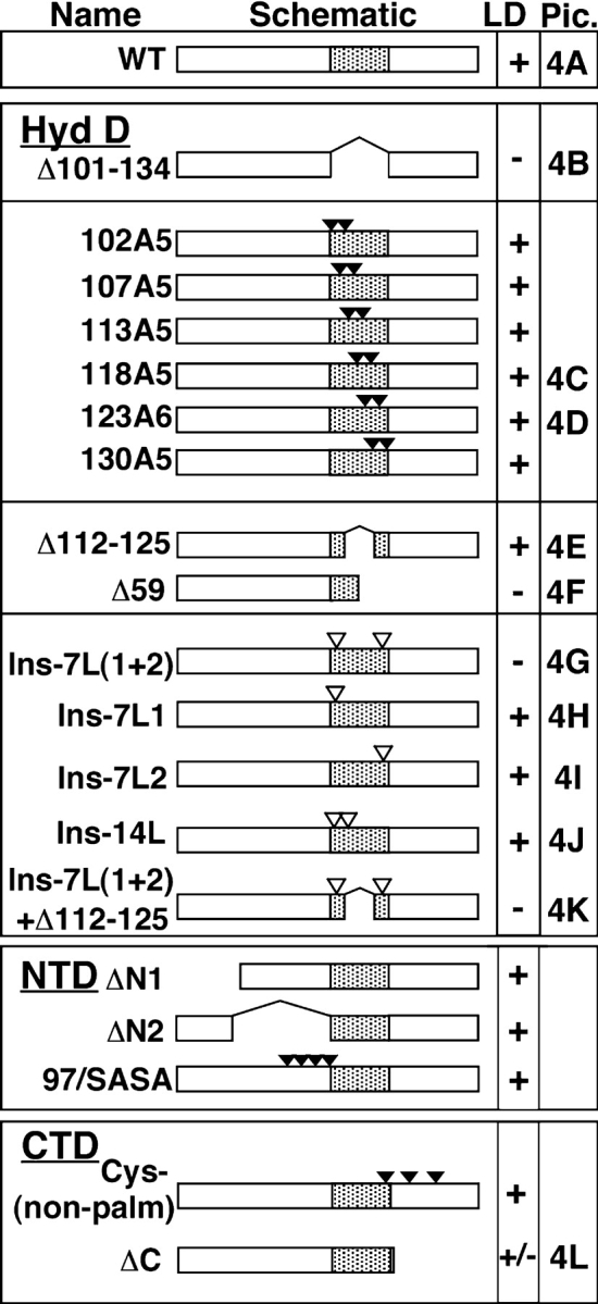
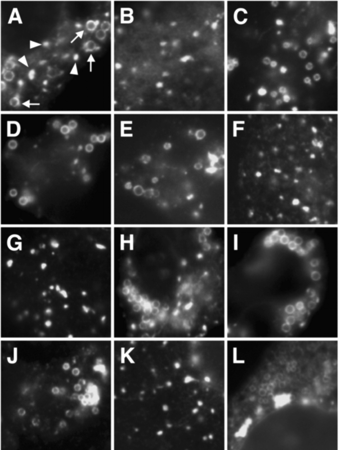
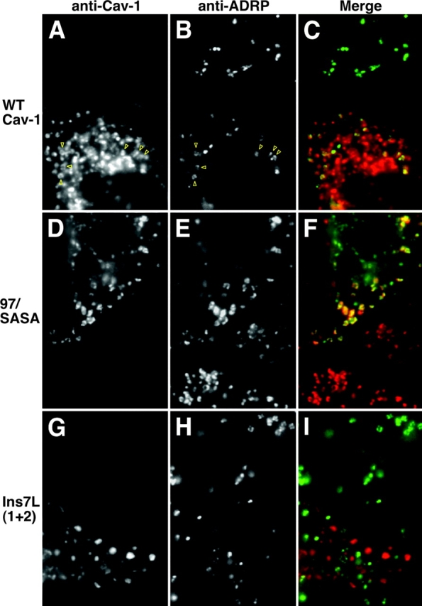
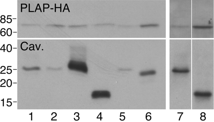
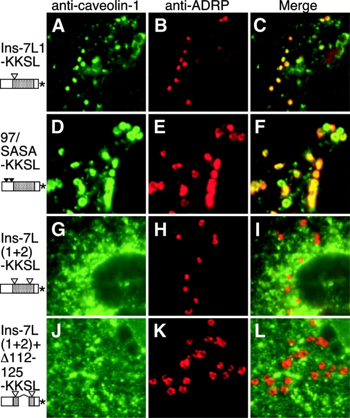
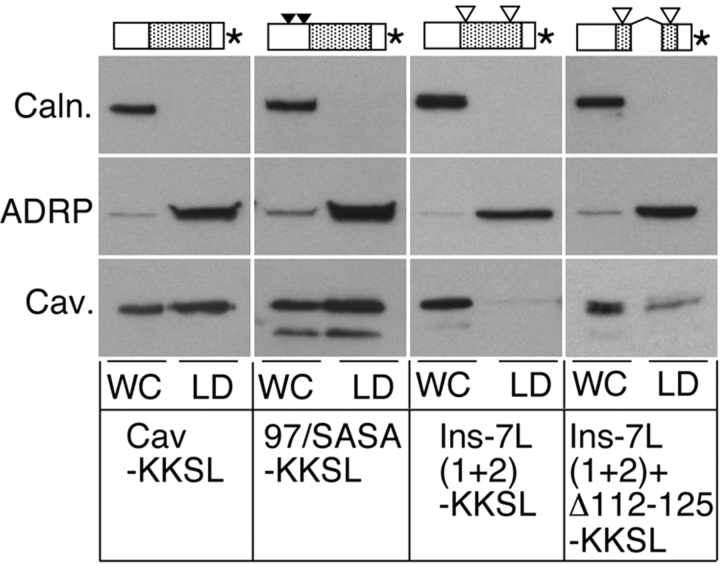
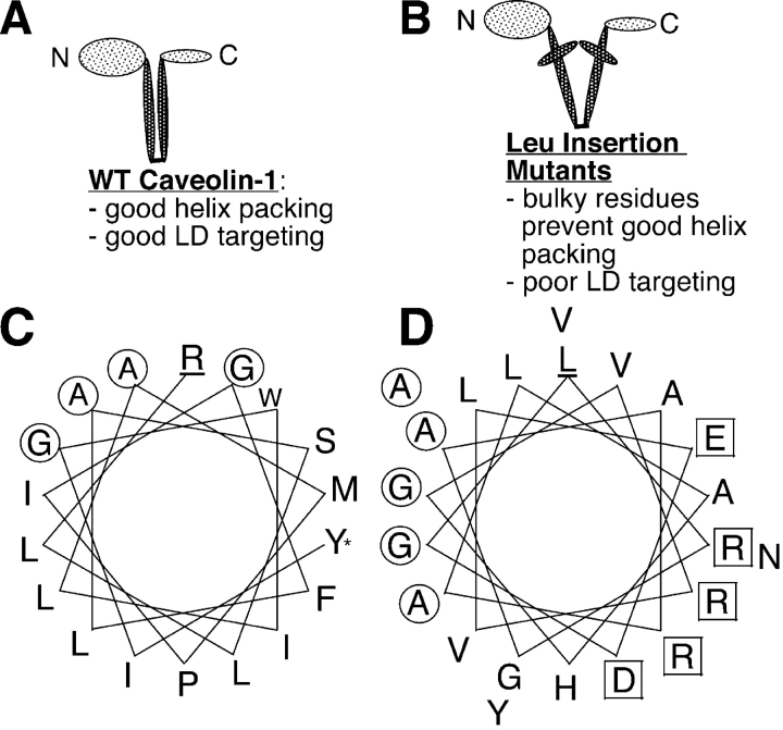
Although caveolins normally reside in caveolae, they can accumulate on the surface of cytoplasmic lipid droplets (LDs). Here, we first provided support for our model that overaccumulation of caveolins in the endoplasmic reticulum (ER) diverts the proteins to nascent LDs budding from the ER. Next, we found that a mutant H-Ras, present on the cytoplasmic surface of the ER but lacking a hydrophobic peptide domain, did not accumulate on LDs. We used the fact that wild-type caveolin-1 accumulates in LDs after brefeldin A treatment or when linked to an ER retrieval motif to search for mutants defective in LD targeting. The hydrophobic domain, but no specific sequence therein, was required for LD targeting of caveolin-1. Certain Leu insertions blocked LD targeting, independently of hydrophobic domain length, but dependent on their position in the domain. We propose that proper packing of putative hydrophobic helices may be required for LD targeting of caveolin-1.


Figures

Figure 1.
Figure 1.
Effect of drugs on LD accumulation of caveolin-1. COS cells were left untreated (A–C) or treated with BFA alone (D–F) or BFA with nocodazole (G–I), cytochalasin B (J–L), or cycloheximide (M–O) for 5 h. Caveolin-1 was detected by IF with anti–caveolin-1 and Alexa Fluor 350-GAR (left) and LDs with Nile red (center). Right, merged images. Images were taken with a 100× objective. Each image shows a portion of one cell; for orientation, dark areas at lower right in F and I are the edges of nuclei. Bar, 5 μm.
Figure 2.
Figure 2.
Neither H-Ras nor the transmembrane protein PLAP-HA accumulate in LDs. (A, B, D, and E) GFP-tagged nonpalmitoylated H-Ras (A and B) and Cav-KKSL (D and E) were coexpressed in COS cells. Cells were not treated (A and D; same cell) or treated (B and E; same cell) with BFA for 5 h. Mutant H-Ras was visualized by GFP fluorescence, and caveolin-KKSL by IF, using Texas red–GAR. (C and F) In one cotransfected, BFA-treated FRT cell, PLAP-HA was detected with anti-PLAP and FITC-GAR (C), and Cav-KKSL was detected with anti-myc and Texas red–GAM (F).
Figure 3.
Figure 3.
BFA-induced LD accumulation of wild-type and mutant caveolin-1. FRT cells expressing the indicated proteins were treated with BFA for 5 h, and caveolin-1 was detected by IF. Proteins are listed by name and diagrammed schematically (not to scale). The NH2-terminal, hydrophobic, and COOH-terminal domains of caveolin-1 are schematized as open, shaded, and open boxes, respectively. Deletions are schematized as gaps, substitutions as closed triangles, and 7-Leu insertions as open triangles. Except for the hydrophobic domain mutants (102A5–130A5), the number of triangles corresponds to the number of changes. Mutants in the hydrophobic domain (Hyd D), NH2-terminal domain (NTD), and COOH-terminal domain (CTD) are grouped together. Pic., IF images of these proteins are shown in the indicated panels of Fig. 4.
Figure 4.
Figure 4.
Localization of wild-type and mutant caveolin-1 in BFA-treated FRT cells. (A) Caveolin-1; (B) Δ101-134; (C) 118A5; (D) 123A6; (E) Δ112-125; (F) Δ59; (G) Ins7L(1+2); (H) Ins-7L1; (I) Ins-7L2; (J) Ins-14L; (K) Ins7L(1+2)+ Δ112-125; and (L) ΔC. Cells were treated with BFA for 5 h. Proteins were visualized by IF, using anticaveolin antibodies and DTAF-GAR. Arrows, LDs. Arrowheads, puncta staining for GM130 (not depicted), presumed to be ER exit sites.
Figure 5.
Figure 5.
Localization of wild-type and mutant caveolin-1 and ADRP in BFA-treated FRT cells. FRT cells expressing caveolin-1 (A–C), 97/SASA (D–F), or Ins-7L(1+2) (G–I) were BFA treated for 4 h and methanol fixed. Caveolin and mutants were detected by IF, using (A and G) Texas red–GAR or (D) FITC-GAR. ADRP was detected in the transfected cells and neighboring untransfected cells using (B and H) DTAF-GAM or (E) Texas red–GAM. (C, F, and I) Merged images. (A and B) Yellow arrows indicate some regions of overlap.
Figure 6.
Figure 6.
Expression level of caveolin-1 mutants does not correlate with LD targeting. FRT cells were cotransfected with PLAP-HA (as a transfection and loading control) and either Ins-7L1 (lane 1), Ins-7L2 (lane 2), Ins-7L(1+2)+Δ112-125 (lane 3), Δ59 (lane 4), Ins-7L(1+2) (lane 5), or Δ112-125 (lane 6); or, in a separate experiment, analyzed on a separate gel with Ins-7L(1+2) (lane 7) or ΔC (lane 8). Cells were lysed with gel loading buffer and proteins in equal aliquots of lysate were analyzed by SDS-PAGE and Western blotting. Blots were cut, and the tops were probed with anti-PLAP antibodies and the bottoms with anticaveolin antibodies. Both halves were probed with HRP–donkey anti–rabbit IgG, and proteins were visualized by ECL.
Figure 7.
Figure 7.
Localization of KKSL-tagged caveolin-1 mutants. FRT cells expressing Ins-7L1-KKSL (A–C), 97/SASA-KKSL (D–F), Ins-7L(1+2)-KKSL (G–I), or Ins-7L(1+2)+Δ112-125-KKSL (J–L) were methanol fixed. Caveolin-1 proteins (A, D, G, and J) and ADRP in the same cells (B, E, H, and K) were detected by IF. (C, F, I, and L) Merged images. Schematic depictions of proteins are as in Fig. 3, except that only two triangles indicate the four substitutions in 97/SASA. *, KKSL tag.
Figure 8.
Figure 8.
KKSL-tagged caveolin-1 mutants in isolated LDs. LDs were isolated from oleic acid–treated FRT cells expressing Cav-KKSL, 97/SASA-KKSL, Ins-7L(1+2)-KKSL, or Ins-7L(1+2)+Δ112-125-KKSL as indicated. Proteins in the LDs (LD) or in a reserved 5% of the whole cell homogenate (WC) were analyzed by SDS-PAGE and Western blotting and detected by ECL. Blots were cut in three parts. Top, probed with anticalnexin and HRP-donkey anti–rabbit IgG. Middle, anti-ADRP and HRP-GAM. Bottom, anticaveolin and HRP-donkey anti–rabbit IgG. Schematic depictions of proteins are as in Fig. 7.
Figure 9.
Figure 9.
Model depicting role of hydrophobic helix packing in LD targeting of caveolin-1. We speculate here that the hydrophobic domain of caveolin-1 forms two α helices separated by a tight turn. (A) We propose that correct packing of the putative hydrophobic helices is required for LD targeting of wild-type caveolin-1. (B) Insertion of bulky Leu residues in both helices could inhibit LD targeting sterically by preventing proper helix packing. (C) Helical wheel depiction of the first half of the hydrophobic domain of caveolin-1 (R101 at the membrane interface [underlined] through Y119 [*]). Circles, residues on a Gly + Ala–rich helix face. (D) L144 (underlined) through A165 of the core coding region of HCV strain Glasgow (genotype 1a; Hope and McLauchlan, 2000), just downstream of the P138 + P143 motif, modeled as an α helix. Circles, residues on a Gly + Ala–rich helix face. Squares, charged residues.

Similar articles

Cited by

References

    1. Abell, B.M., I.A. Holbrook, M. Abenes, D.J. Murphy, M.J. Hills, and M.M. Moloney. 1997. Role of the proline knot motif in oleosin endoplasmic reticulum topology and oil body targeting. Plant Cell. 9:1481–1493. - PMC - PubMed
    1. Abell, B.M., S. High, and M.M. Moloney. 2002. Membrane topology of oleosin is constrained by its long hydrophobic domain. J. Biol. Chem. 277:8602–8610. - PubMed
    1. Arbuzova, A., L. Wang, J. Wang, G. Hangyas-Mihalyne, D. Murray, B. Honig, and S. McLaughlin. 2000. Membrane binding of peptides containing both basic and aromatic residues. Experimental studies with peptides corresponding to the scaffolding region of caveolin and the effector region of MARCKS. Biochemistry. 39:10330–10339. - PubMed
    1. Arreaza, G., and D.A. Brown. 1995. Sorting and intracellular trafficking of a glycosylphosphatidylinositol-anchored protein and two hybrid proteins with the same ectodomain in MDCK kidney epithelial cells. J. Biol. Chem. 270:23641–23647. - PubMed
    1. Barba, G., F. Harper, T. Harada, M. Kohara, S. Goulinet, Y. Matsuura, G. Eder, Z. Schaf, M.J. Chapman, T. Miyamura, and C. Brechot. 1997. Hepatitis C virus core protein shows a cytoplasmic localization and associates to cellular lipid storage droplets. Proc. Natl. Acad. Sci. USA. 94:1200–1205. - PMC - PubMed

Publication types

MeSH terms