. 2004 Feb 1;117(Pt 4):559-70.
doi: 10.1242/jcs.00893. Epub 2004 Jan 6.

Mechanism of recruiting Sec6/8 (exocyst) complex to the apical junctional complex during polarization of epithelial cells

Affiliations

Mechanism of recruiting Sec6/8 (exocyst) complex to the apical junctional complex during polarization of epithelial cells

Charles Yeaman et al. J Cell Sci. .

Abstract

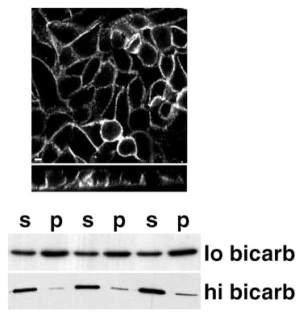
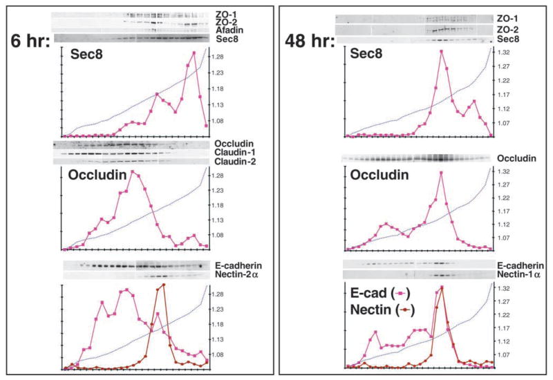
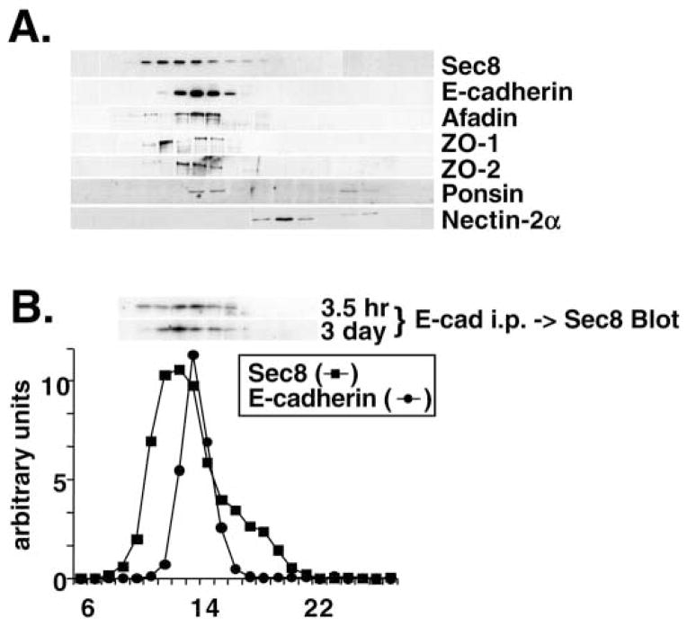
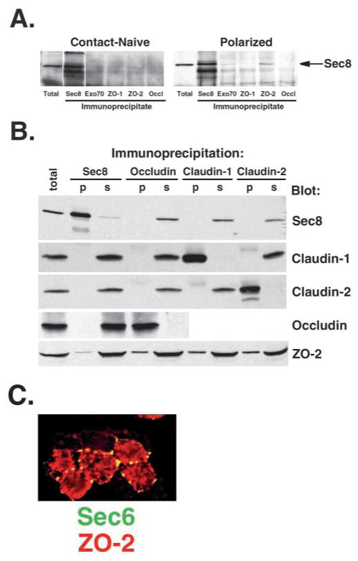
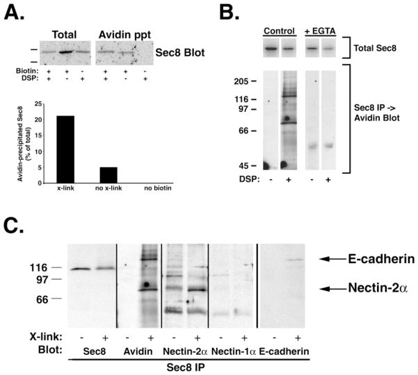
Sec6/8 (exocyst) complex regulates vesicle delivery and polarized membrane growth in a variety of cells, but mechanisms regulating Sec6/8 localization are unknown. In epithelial cells, Sec6/8 complex is recruited to cell-cell contacts with a mixture of junctional proteins, but then sorts out to the apex of the lateral membrane with components of tight junction and nectin complexes. Sec6/8 complex fractionates in a high molecular mass complex with tight junction proteins and a portion of E-cadherin, and co-immunoprecipitates with cell surface-labeled E-cadherin and nectin-2alpha. Recruitment of Sec6/8 complex to cell-cell contacts can be achieved in fibroblasts when E-cadherin and nectin-2alpha are co-expressed. These results support a model in which localized recruitment of Sec6/8 complex to the plasma membrane by specific cell-cell adhesion complexes defines a site for vesicle delivery and polarized membrane growth during development of epithelial cell polarity.


Figures

Fig. 1
Fig. 1
Sec6/8 complex becomes restricted to apical junctional complex during development of cell polarity. Contact-naive MDCK cells were seeded at confluent density on collagen-coated filters, allowed to attach in low calcium medium for 3 hours, and then switched to high calcium medium for 0, 1, 3, 6, 12, or 24 hours. At each time point, cultures were fixed with 4% paraformaldehyde and extracted with buffer containing 1% Triton X-100. (A,B) Sec6 distribution was compared to that of either E-cadherin (A) or ZO-1 (B). Anti-Sec6 monoclonal antibody (9H5) was visualized with FITC-labeled goat anti-mouse antibody. Rabbit polyclonal antibodies to E-cadherin and ZO-1 were visualized with Texas Red-labeled donkey anti-rabbit antibody. Confocal images in the upper panels were acquired along the xy axis (en face view) of the cell monolayer. The xz views, in the lower panels, were constructed by averaging sections over a line at each z position in 0.2 μm steps. Scale bar: 10 μm. (C) Relative pixel intensities of Sec6, E-cadherin and ZO-1 fluorescence at each optical section (1=apical, 19=basal) were averaged from five independently scanned fields.
Fig. 2
Fig. 2
Sec6 co-localizes with components of the nectin complex in early cell-cell contacts and polarized MDCK cells. (Top panels) Low-density cultures of MDCK cells were allowed to form calcium-dependent cell-cell contacts for 1 hour, and then fixed with 2% paraformaldehyde before extraction with 1% Triton X-100. Sec6 distribution was compared to that of afadin or nectin-2α. (Bottom panel) Confluent MDCK cultures on polycarbonate filters were fixed and extracted 24 hours after induction of calcium-dependent cell-cell adhesion. Sec6 distribution was compared to that of afadin. Anti-Sec6 monoclonal antibody (9H5) was visualized with FITC-labeled goat anti-mouse antibody (in the afadin panels) or with Texas Red-labeled donkey anti-mouse antibody (in the nectin panel). Rabbit polyclonal antibody to afadin was visualized with Texas Red-labeled donkey anti-rabbit antibody, and rat monoclonal antibody to nectin-2α was visualized with FITC-labeled goat anti-rat antibody. Confocal images were obtained as described in Fig. 1 legend. Scale bar: 10 μm.
Fig. 3
Fig. 3
Effect of sodium bicarbonate concentration on Sec6/8 complex distribution. Confluent MDCK cultures on polycarbonate filters were grown in DMEM containing either 1 g/l (‘lo bicarb’) or 3.7 g/l (‘hi bicarb’) sodium bicarbonate for 48 hours. (Top) Cultures were fixed with 4% paraformaldehyde before extraction with buffer containing 1% Triton X-100. Anti-Sec6 monoclonal antibody (9H5) was visualized with FITC-labeled goat anti-mouse antibody. Confocal images were obtained as described in Fig. 1 legend. Scale bar: 5 μm. (Bottom) Triplicate filters of cells grown in hi or lo bicarbonate were extracted successively in Triton X-100 and SDS, as described in Materials and Methods. Sec8 in Triton-soluble (‘s’) and Triton-insoluble (‘p’) fractions was quantified by SDS-PAGE and western blotting. Protein levels were quantified using a Molecular Dynamics Phosphorimager. In 1 g/l bicarbonate, Sec8 is enriched at the apical junction (Fig. 1) and is only partially (~30%) soluble in Triton X-100. In 3.7 g/l bicarbonate, Sec8 is diffusely distributed along the lateral and basal membranes and is almost entirely (~90%) soluble in Triton X-100.
Fig. 4
Fig. 4
Fractionation of MDCK cells in iodixanol gradients. MDCK cells were homogenized either 6 hours or 48 hours after induction of calcium-dependent cell-cell adhesion. Post-nuclear supernatants were mixed with 10%, 20% and 30% (w/v) iodixanol, layered step-wise in centrifuge tubes and centrifuged at 350,000 g for 3 hours. The presence of Sec8, ZO-1, ZO-2, afadin, occludin, claudin-1, claudin-2, E-cadherin, nectin-1α and nectin-2α in gradient fractions was assayed by SDS-PAGE followed by immunoblotting with specific antibodies. Protein levels were quantified using a Molecular Dynamics Phosphorimager. Densities of each fraction were calculated after measuring refractive indices with a refractometer, and are plotted as dotted lines on each graph with values (in g/ml) indicated on the y-axis.
Fig. 5
Fig. 5
Fractionation of junctional proteins associated with Sec6/8 complex. (A) Detergent extracts of polarized MDCK cells were fractionated by Superose 6 FPLC as described in Materials and Methods. Fractions 6–28 were divided into equal aliquots, separated by SDS-PAGE, and transferred to Immobilon P membranes. Membranes were probed with antibodies to Sec8, E-cadherin, afadin, ZO-1, ZO-2, ponsin or nectin-2α. Protein levels were quantified using a Molecular Dynamics Phosphorimager. The elution profiles of Sec6 and E-cadherin are shown in B. The elution peaks of globular protein standards with known relative molecular masses were also determined: thyroglobulin, Mr=669,000 (fraction 16); apoferritin, Mr=443,000 (fraction 19); catalase, Mr=232,000 (fraction 22); bovine serum albumin, Mr=66,000 (fraction 24). (B) Coimmunoprecipitation of Sec8 with E-cadherin adhesion complex. MDCK cells were extracted either 3.5 hours or 3 days after induction of calcium-dependent cell-cell adhesion and extracts were fractionated by Superose 6 FPLC. Each fraction (10–19) was subjected to immunoprecipitation with anti-E-cadherin E2 antiserum. Immunoprecipitated material was eluted in SDS-PAGE sample buffer and the presence of Sec8 in each fraction was assayed by SDS-PAGE followed by immunoblotting. Protein levels were quantified using a Molecular Dynamics Phosphorimager.
Fig. 6
Fig. 6
Sec8 associates with a fraction of ZO-2. (A) MDCK cells were extracted in 1% Triton X-100 either 0 hours (contact-naive) or 48 hours (polarized) after inducing calcium-dependent cell-cell adhesion. Extracts were subjected to immunoprecipitation with specific antibodies to Sec8, Exo70, ZO-1, ZO-2 or occludin. The presence of Sec8 in precipitated immune complexes was assessed by SDS-PAGE followed by immunoblotting with Sec8 antibodies. (B) MDCK cells were homogenized 48 hours after induction of calcium-dependent cell-cell adhesion and junction-enriched membrane fractions were isolated by isopycnic density gradient centrifugation as described in Fig. 2. Membranes were extracted in 1% Triton X-100 and subjected to immunoprecipitation with antibodies specific for Sec8, occludin, claudin-1 or claudin-2. The presence of each of these proteins and of ZO-2 in precipitated immune complexes was assessed by SDS-PAGE followed by immunoblotting with specific antibodies. (C) Polarized MDCK cultures on polycarbonate filters were treated with 2 μM latrunculin B for 1 hour, then fixed with 2% paraformaldehyde before extraction with buffer containing 1% Triton X-100. Anti-Sec6 monoclonal antibody (9H5) was visualized with FITC-labeled goat anti-mouse antibody and anti-ZO-2 polyclonal antibody was visualized with Texas Red-labeled donkey anti-rabbit antibody.
Fig. 7
Fig. 7
Sec6/8 complex is associated with E-cadherin and nectin-2α. Polarized MDCK cells cultured on polycarbonate filters were surface labeled with either Sulfo-NHS-SS-Biotin (A) or Sulfo-NHS-LC-LC-Biotin (B,C) and extracted with 1% Triton X-100 either directly (no x-link or –DSP) or following (x-link or +DSP) chemical cross-linking with the membrane-permeable cross-linker DSP. Non-biotinylated controls (no biotin) were subjected to cross-linking prior to extraction. (A) Extracts were incubated with avidin-agarose, and the presence of Sec8 in avidin precipitates was assayed by SDS-PAGE followed by immunoblotting with anti-Sec8 antibody. (B,C) Extracts were subjected to immunoprecipitation with anti-Sec8 antibodies, and the presence of biotinylated proteins, Sec8, nectin-2α, nectin-1α and E-cadherin in precipitated immune complexes was assessed by SDS-PAGE followed by immunoblotting with HRP-avidin or specific antibodies. ‘+EGTA’ cultures were incubated in LCM + 2 mM EGTA for 6 hours prior to biotinylation.
Fig. 8
Fig. 8
E-cadherin and nectin-2α cooperate to recruit Sec6/8 complex to intercellular contacts in fibroblasts. LE cells, or LE cells that had been transiently transfected with pcDNA3.1-IgK-2HA-nectin-2α, were cultured without (no Dex) or with (plus Dex) 10−6 M dexamethasone for 18 hours to induce E-cadherin expression. Cells were fixed with 4% paraformaldehyde and then extracted with buffer containing 1% Triton X-100. Anti-Sec8 monoclonal antibody (2E9) was visualized either with FITC- or Texas Red-labeled secondary antibodies. Anti-E-cadherin polyclonal antibody (UVO) was visualized with Texas Red-labeled secondary antibody. Anti-nectin-2α rat monoclonal antibody was visualized with FITC-labeled secondary antibody. Arrows indicate homotypic cell-cell contacts formed between adjacent cells expressing either nectin-2α or E-cadherin only. Arrowheads indicate cell-cell contacts between E-cadherin-expressing cells in which nectin-2α was also expressed. Scale bar: 30 μm.

Similar articles

Cited by

References

    1. Ando-Akatsuka Y, Yonemura S, Itoh M, Furuse M, Tsukita S. Differential behavior of E-cadherin and occludin in their colocalization with ZO-1 during the establishment of epithelial cell polarity. J Cell Physiol. 1999;179:115–125. - PubMed
    1. Angres B, Barth A, Nelson WJ. Mechanism for transition from initial to stable cell-cell adhesion: kinetic analysis of E-cadherin-mediated adhesion using a quantitative adhesion assay. J Cell Biol. 1996;134:549–557. - PMC - PubMed
    1. Aoki J, Koike S, Asou H, Ise I, Suwa H, Tanaka T, Miyasaka M, Nomoto A. Mouse homolog of poliovirus receptor-related gene 2 product, mPRR2, mediates homophilic cell aggregation. Exp Cell Res. 1997;235:374–384. - PubMed
    1. Ausubel FM, Brent R, Kingston RE, Moore DD, Seidman JG, Smith JA, Struhl K. Current Protocols in Molecular Biology. New York: John Wiley and Sons; 1987.
    1. Bagnat M, Chang A, Simons K. Plasma membrane proton ATPase Pma1p requires raft association for surface delivery in yeast. Mol Biol Cell. 2001;12:4129–4138. - PMC - PubMed

Publication types