

doi: 10.1038/sj.embor.7400056.
Life in a crowded world
Affiliations
- PMID: 14710181
- PMCID: PMC1298967
- DOI: 10.1038/sj.embor.7400056
Item in Clipboard
Life in a crowded world
EMBO Rep.
2004 Jan.
Abstract
Workshop on the Biological Implications of Macromolecular Crowding
Figures

Excluded volume (shown in pink and black) and available volume (shown in blue) in a solution of background macromolecules. The two panels show the volume available to the centre of a test molecule (T) either (A) much smaller than or (B) of similar size to the background macromolecules (see the text for details). Reproduced with permission from Minton (2001). (Figure presented at the workshop by A. Minton.)

Calculated change in the equilibrium constant for heteroassociation of dilute globular protein A (65 kDa) and dilute globular protein B (52 kDa) as a function of the fraction of volume occupied by a 27-kDa inert protein (upper curve) and by a 70-kDa inert protein (lower curve). All proteins are represented by equivalent hard spherical particles with respective radii proportional to the cube root of molar mass; calculations were performed with the scaled particle theory of hard sphere mixtures (Lebowitz et al, 1965). For details of the calculation see Minton (1998).

Cell compartments are crowded. Shown is a three-dimensional reconstruction of part of the cytoplasm of a Dictyostelium discoideum cell, produced by the Baumeister group (Medalia et al, 2002). Actin filaments are shown in orange, ribosomes and other macromolecular assemblies in grey, and membrane structures in blue. (Figure presented at the workshop by S. Nickel.)
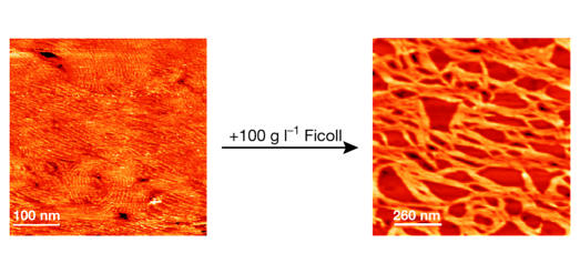
Crowding-induced formation of ribbons of the bacterial cell division FtsZ protein as monitored by atomic force microscopy. The left-hand image shows the thin FtsZ filaments observed in vitro in the absence of crowding. The right-hand image shows the FtsZ ribbons formed after the addition of 100 g l−1 Ficoll 70, a 70-kDa inert crowder. (Figure presented at the workshop by J. González; see González et al, 2003).

This workshop took place at the Palacio de Magalia (Las Navas del Marqués, Avila, Spain), between 14 and 18 June 2003, and was organized by J. Ellis, A. Minton and G. Rivas. Further details on the workshop can be found at http://www.cib.csic.es/~revers/embo2003/index.htm


References
-
- Baumeister W (2002) Electron tomography: towards visualizing the molecular organization of the cytoplasm. Curr Opin Struct Biol 12: 679–684 - PubMed
-
- Bray D (1998) Signalling complexes: biophysical constraints on intercellular communication. Annu Rev Biophys Biomol Struct 27: 59–75 - PubMed
-
- Brown MP, Royer C (1997) Fluorescence spectroscopy as a tool to investigate protein interactions. Curr Opin Biotech 8: 45–49 - PubMed
Publication types
MeSH terms
Substances
LinkOut - more resources
Full Text Sources
Other Literature Sources
