Comparative Study
. 2004 Jan;5(1):41-6.
doi: 10.1038/sj.embor.7400050.

A test of the CoHR motif associated with meiotic double-strand breaks in Saccharomyces cerevisiae

Affiliations
Comparative Study

A test of the CoHR motif associated with meiotic double-strand breaks in Saccharomyces cerevisiae

Stuart J Haring et al. EMBO Rep. 2004 Jan.

Abstract

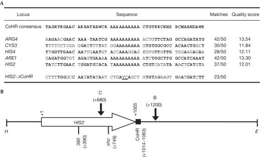
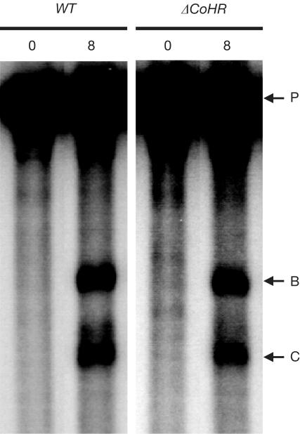
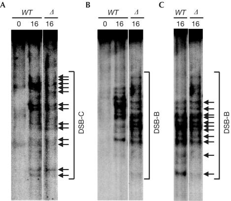
Meiotic recombination is not random along chromosomes; rather, there are preferred regions for initiation called hotspots. Although the general properties of meiotic hotspots are known, the requirements at the DNA sequence level for the determination of hotspot activity are still unclear. The sequence of six known hotspots in Saccharomyces cerevisiae was compared to identify a common homology region (CoHR). They reported that the locations of CoHR sequences correspond to mapped double-strand break (DSB) sites along three chromosomes (I, III, VI). We report here that a deletion of CoHR at HIS2, a hotspot used to identify the motif, has no significant effect on recombination. In the absence of CoHR, DSB formation occurs at a high frequency and at the same sequences as in wild-type strains. In cases where the deletion of sequences containing the CoHR motif has been shown to reduce recombination, we propose that it may be a reflection of the location of the deletion, rather than the loss of CoHR, per se.


Figures

Figure 1
Figure 1
CoHR sequences and features at HIS2. (A) Comparison of the CoHR sequences of five known meiotic recombination hotspots (modified from Fig 1 of Blumental-Perry et al, 2000) and the sequence created by deleting CoHR at HIS2. Sequences and base designations are as in Fig 1 from Blumental-Perry et al (2000), except that the poly(A) region has been corrected from 12 to 10 bp. Matches (in bold) represent base pairs matching the CoHR consensus. The quality score has been taken from Table 1 of Blumental-Perry et al (2000). A comparison of the fusion created by ΔCoHR with the CoHR consensus is shown. Underlined bases represent the fusion junction created by ΔCoHR. (B) Important features at HIS2. The 2.6 kbp HindIII–EcoRI fragment containing HIS2 is shown. The direction of transcription of HIS2 is designated by the horizontal arrow. DSB sites are shown by vertical arrows, with C and B representing DSB-C and DSB-B, respectively. The location of CoHR is designated by the solid box at the end of the HIS2 coding region. Alleles used to measure gene conversion are designated by 390 and xho (Malone et al, 1992, 1994). E=EcoRI; H=HindIII.
Figure 2
Figure 2
Relationships between CoHR sites and DSB sites on chromosome III. (A) Distribution of the distance between CoHR sites and the nearest DSB sites. The actual distribution, determined by combining data from Baudat & Nicolas (1997) and Blumental-Perry et al (2000), is represented by black bars. The random distribution determined by 1,000 independent trials (see Methods) is represented by grey bars. (B) Plot of DSB strength versus distance to nearest CoHR. All detectable DSBs within 4.36 kbp of a CoHR motif were examined. (C) Plot of DSB strength nearest to each CoHR versus CoHR quality score. (D) Plot of DSB strength nearest to each CoHR versus CoHR quality score, as in (C), excluding the ARE1 data point.
Figure 3
Figure 3
DSB formation at HIS2 in the presence and absence of the CoHR site. WT refers to a diploid in which CoHR is intact at HIS2; ΔCoHR refers to a diploid in which CoHR is deleted at HIS2. The numbers above each lane represent hours in sporulation. DSB-B, DSB-C and the parental (unbroken) fragments are designated by horizontal arrows labelled B, C and P, respectively.
Figure 4
Figure 4
Medium-resolution mapping of DSBs at HIS2 in the presence and absence of the CoHR site. DSBs at indistinguishable sequences in wild-type or ΔCoHR strains are designated by horizontal arrows. WT refers to a wild-type diploid with CoHR intact at HIS2; Δ refers to a diploid with CoHR deleted at HIS2. Numbers above lanes represent hours in sporulation. (A) Mapping of the break pattern at DSB-C. (B, C) Mapping of the break pattern at DSB-B. The pattern of DSB-B in (B) appears altered in a ΔCoHR strain, because the DSB fragments also contain the ΔCoHR (50 bp) deletion. To account for the 50 bp size difference due to ΔCoHR, the WT lane from (B) was stretched in (C) to mimic the running of 50 bp shorter fragments on a gel.

Similar articles

Cited by

References

    1. Alani E, Padmore R, Kleckner N (1990) Analysis of wild-type and rad50 mutants of yeast suggests an intimate relationship between meiotic chromosome synapsis and recombination. Cell 61: 419–436 - PubMed
    1. Baker BS, Carpenter AT, Esposito MS, Esposito RE, Sandler L (1976) The genetic control of meiosis. Annu Rev Genet 10: 53–134 - PubMed
    1. Baudat F, Nicolas A (1997) Clustering of meiotic double-strand breaks on yeast chromosome III. Proc Natl Acad Sci USA 94: 5213–5218 - PMC - PubMed
    1. Blumental-Perry A, Zenvirth D, Klein S, Onn I, Simchen G (2000) DNA motif associated with meiotic double-strand break regions in Saccharomyces cerevisiae. EMBO Rep 1: 232–238 - PMC - PubMed
    1. Borde V, Wu TC, Lichten M (1999) Use of a recombination reporter insert to define meiotic recombination domains on chromosome III of Saccharomyces cerevisiae. Mol Cell Biol 19: 4832–4842 - PMC - PubMed

Publication types

LinkOut - more resources