Comparative Study
. 2004 Jan;5(1):71-7.
doi: 10.1038/sj.embor.7400036.

Reconsidering the evolution of eukaryotic selenoproteins: a novel nonmammalian family with scattered phylogenetic distribution

Affiliations
Comparative Study

Reconsidering the evolution of eukaryotic selenoproteins: a novel nonmammalian family with scattered phylogenetic distribution

Sergi Castellano et al. EMBO Rep. 2004 Jan.

Abstract

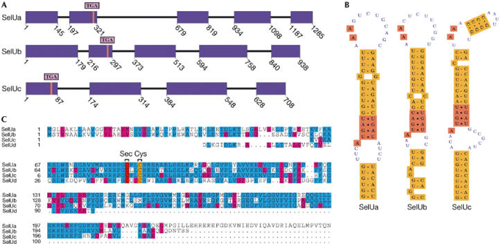
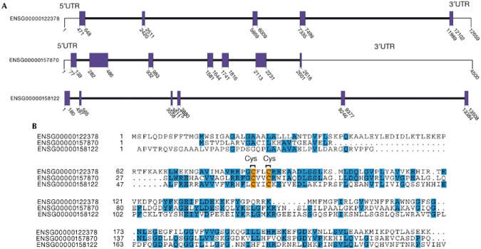
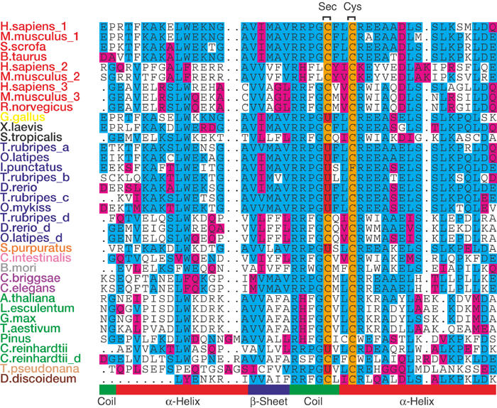
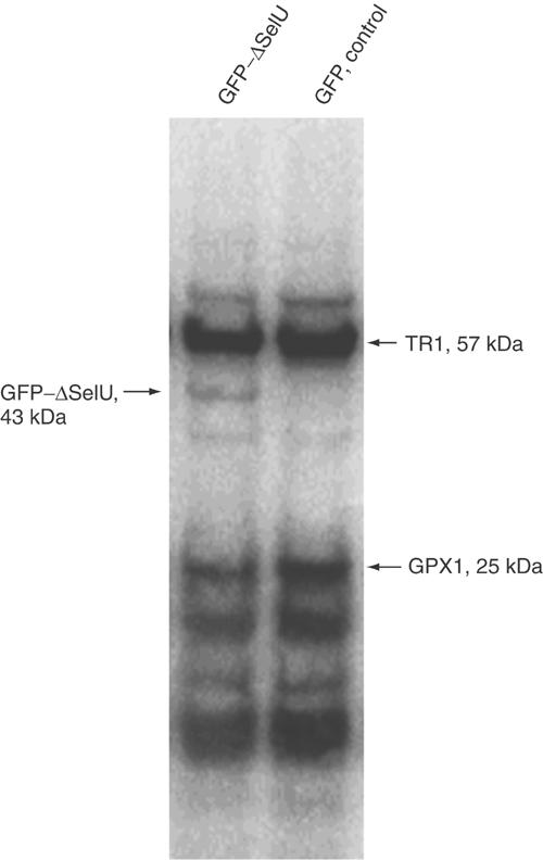
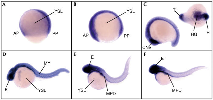
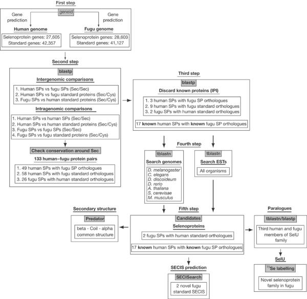
While the genome sequence and gene content are available for an increasing number of organisms, eukaryotic selenoproteins remain poorly characterized. The dual role of the UGA codon confounds the identification of novel selenoprotein genes. Here, we describe a comparative genomics approach that relies on the genome-wide prediction of genes with in-frame TGA codons, and the subsequent comparison of predictions from different genomes, wherein conservation in regions flanking the TGA codon suggests selenocysteine coding function. Application of this method to human and fugu genomes identified a novel selenoprotein family, named SelU, in the puffer fish. The selenocysteine-containing form also occurred in other fish, chicken, sea urchin, green algae and diatoms. In contrast, mammals, worms and land plants contained cysteine homologues. We demonstrated selenium incorporation into chicken SelU and characterized the SelU expression pattern in zebrafish embryos. Our data indicate a scattered evolutionary distribution of selenoproteins in eukaryotes, and suggest that, contrary to the picture emerging from data available so far, other taxa-specific selenoproteins probably exist.


Figures

Figure 1
Figure 1
Fugu SelU family. (A) Gene structure (coding exons in purple) plotted using gff2ps (Abril & Guigó, 2000). Red lines mark the TGA triplet. SelUc is a partial gene lacking the upstream region. (B) SECIS structures. SelUa and SelUb bear a type 1 SECIS and SelUc a type 2 SECIS. (C) Alignment of SelU paralogues using CLUSTAL_W (Thompson et al, 1994). U is Sec.
Figure 2
Figure 2
Ensembl human SelU family. (A) Gene structure (coding exons) for ENSG00000122378, ENSG00000157870 and ENSG00000158122 genes. (B) Alignment of SelU paralogues.
Figure 3
Figure 3
Multiple alignment of SelU proteins across the eukaryotic lineage (the sequence around the Sec (U) amino acid in red and Cys (C) in orange is shown). The sequences are clustered phylogenetically and by sequence similarity. The predicted protein secondary structure is shown at the bottom (also see supplementary information online). Species colours: mammals, red; birds, yellow; amphibians, black; fish, blue; echinoderms, orange; tunicates, pink; arthropods, grey; worms, violet; plants, green; diatoms, light orange; slime molds, brown.
Figure 4
Figure 4
Detection of 75Se-labelled SelU. CV-1 cells were transfected with either GFP–ΔSelU fusion construct (left line) or GFP vector as a control (right line), and grown in the presence of 75Se[selenite] for 24 h. Cell extracts containing 75Se-labelled selenoproteins were resolved by SDS–polyacrylamide gel electrophoresis and visualized with a PhosphorImager System. Locations of major endogenous selenoproteins TR1 (57 kDa) and GPX1 (25 kDa) are shown on the right, and the GFP–ΔSelU fusion protein on the left.
Figure 5
Figure 5
Expression pattern of the SelU gene during development in zebrafish embryos. Developmental stages are (A) gastrula, (B) early somitogenesis, (C) late somitogenesis, (D) 24 h postfertilization, (E) 36 h postfertilization and (F) 48 h postfertilization. All views are lateral except the one in the upper right corner in (C) which is dorsoventral. AP, anterior pole; CNS, central nervous system; E, eye; H, head; HG, hatching gland; MPD, medial part of the pronephric duct; MY, myotomes; PP, posterior pole; T, tail; YSL, yolk syncytial layer.
Figure 6
Figure 6
General schema for selenoprotein identification.

References

    1. Abril JF, Guigó R (2000) gff2ps: visualizing genomic annotations. Bioinformatics 16: 743–744 - PubMed
    1. Altschul SF, Madden T, Schaffer A, Zhang J, Zhang Z, Miller W, Lipman D (1997) Gapped BLAST and PSI-BLAST: a new generation of protein database search programs. Nucleic Acids Res 25: 3389–3402 - PMC - PubMed
    1. Axley MJ, Böck A, Stadman TC (1991) Catalytic properties of an Escherichia coli formate dehydrogenase mutant in which sulfur replaces selenium. Proc Natl Acad Sci USA 88: 8450–8454 - PMC - PubMed
    1. Berry MJ, Mai AL, Kieffer J, Harney JW, Larsen P (1992) Substitution of cysteine for selenocysteine in type i iodothyronine deiodinase reduces the catalytic efficiency of the protein but enhances its translation. Endocrinology 131: 1848–1852 - PubMed
    1. Castellano S, Morozova N, Morey M, Berry MJ, Serras F, Corominas M, Guigó R (2001) In silico identification of novel selenoproteins in the Drosophila melanogaster genome. EMBO Rep 2: 697–702 - PMC - PubMed

Publication types