. 2004 Jan 12;90(1):263-9.
doi: 10.1038/sj.bjc.6601473.

Encapsulation into sterically stabilised liposomes enhances the immunogenicity of melanoma-associated Melan-A/MART-1 epitopes

Affiliations

Encapsulation into sterically stabilised liposomes enhances the immunogenicity of melanoma-associated Melan-A/MART-1 epitopes

M Adamina et al. Br J Cancer. .

Abstract

Tumour-associated antigens (TAA)-specific vaccination requires highly immunogenic reagents capable of inducing cytotoxic T cells (CTL). Soluble peptides are currently used in clinical applications despite an acknowledged poor immunogenicity. Encapsulation into liposomes has been suggested to improve the immunogenicity of discrete antigen formulations. We comparatively evaluated the capacity of HLA-A2.1 restricted Melan-A/MART-1 epitopes in soluble form (S) or following inclusion into sterically stabilised liposomes (SSL) to be recognised by specific CTL, to stimulate their proliferation and to induce them in healthy donors' peripheral blood mononuclear cells (PBMC), as well as in melanoma-derived tumour-infiltrating lymphocytes (TIL). HLA-A2.1(+), Melan-A/MART-1-NA-8 melanoma cells served as targets of specific CTL in 51Cr release assays upon pulsing by untreated or human plasma-treated soluble or SSL-encapsulated Melan-A/MART-1 27-35 (M27-35) or 26-35 (M26-35) epitopes. These reagents were also used to stimulate CTL proliferation, measured as 3H-thymidine incorporation, in the presence of immature dendritic cells (iDC), as antigen-presenting cells (APC). Induction of specific CTL upon stimulation with soluble or SSL-encapsulated peptides was attempted in healthy donors' PBMC or melanoma-derived TIL, and monitored by 51Cr release assays and tetramer staining. Na-8 cells pulsing with SSL M27-35 resulted in a five-fold more effective killing by specific CTL as compared with equal amounts of S M27-35. Encapsulation into SSL also provided a partial (50%) protection of M27-35 from plasma hydrolysis. No specific advantages regarding M26-35 were detectable in these assays. However, at low epitope concentrations (</=100 ng ml(-1)), SSL M26-35 was significantly more effective in inducing CTL proliferation than S M26-35, in the presence of iDC, as APC. Preincubation with iDC for 6 h virtually abolished the capacity of S M26-35 to stimulate specific CTL proliferation, but only partially affected that of SSL M26-35. Most importantly, SSL M26-35 was able to enhance the induction of specific CTL in healthy donors PBMC and in melanoma-derived TIL as compared to S M26-35. Taken together, our data indicate that encapsulation of TAA epitopes into SSL results in effective immunogenic formulations suitable for clinical use in active specific tumour immunotherapy.


Figures

Figure 1
Figure 1
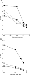
Immunorecognition of Melan-A/MART-1 epitopes by specific CTL. HLA-A2.1-positive, Melan-A/MART-1-negative NA-8 cells were 51Cr labelled and incubated for 2 h in the presence of graded amounts of Melan-A/MART-1 27–35 (M27–35, panel (A)) or 26–35 (M26–35, panel (B)) epitopes or control peptides in soluble form (SC) or encapsulated into sterically stabilised liposomes (SSL C). Cells were then washed and cultured for 4 h in the presence of a specific HLA-A2.1-restricted cytotoxic T-cell clone at 5 : 1 effector target ratio. Supernatants were then harvested and 51Cr release was measured by a gamma counter. Data are reported as mean % specific target cell lysis from triplicate wells. Standard deviations, never exceeding 10% of the reported values, were omitted.
Figure 2
Figure 2
Antigenic peptide degradation in the presence of human plasma. Melan-A/MART-1 27–35 (M27–35, 1 μg ml−1) or 26–35 (M26–35, 8 ng ml−1) peptides in soluble form (S) or encapsulated into SSL were incubated for the indicated times at 37°C in the presence of undiluted human plasma. The different preparations were then used to pulse 51Cr-labelled Na-8 cells for 2 h. These were cocultured for 4 h in the presence of a specific HLA-A2.1-restricted cytotoxic T-cell clone at 5 : 1 effector target ratio in triplicate samples. Supernatants were then harvested and 51Cr release was measured by a gamma counter. Data are reported as % of the cytotoxic activity detected in the presence of the corresponding untreated M27–35 or 26–35 preparations. Standard deviations, never exceeding 10% of the reported values, were omitted.
Figure 3
Figure 3
Induction of specific CTL proliferation by Melan-A/MART-1 26–35 peptide: iDC as antigen-presenting cells. (A) Cells from a Melan-A/MART-1-specific, HLA-A2.1-restricted CTL clone were cultured in the presence of irradiated, HLA-A2.1-matched, CD14/CD1a+ immature, monocyte-derived dendritic cells, and the indicated concentrations of Melan-A/MART-1 26–35 or control peptides in soluble form (shaded and white columns, respectively) or contained into SSL (black and striped columns, respectively). Proliferation of triplicate wells was assessed by 3H-thymidine on day three of culture. (B) HLA-A2.1-positive monocyte-derived CD14/CD1a+ iDC were cultured for 3 or 6 h (middle and lower panels, respectively) in the presence of Melan-A/MART-1 26–35 or control peptides in soluble form (shaded and white columns, respectively) or contained into SSL (black and striped columns, respectively) at the indicated concentrations. T cells from a specific CTL clone were then added. Data reported in the upper panel refer to cultures where iDC, CTL and immunogenic materials were simultaneously added without preincubation steps. In all cases, 3H-thymidine incorporation of triplicate wells was measured after 3 days culture.
Figure 4
Figure 4
Induction of Melan-A/MART-1 specific CTL by synthetic peptides encapsulated into SSL. Healthy donor's PBMC were cultured for 1 week in the presence of Melan-A/MART-1 26–35 or control peptides at 10 μg ml−1 in soluble form (shaded symbols) or encapsulated into SSL (black symbols). rIL-2 (20 U ml−1) was then added and cultures were restimulated on day 10 and weekly thereafter with the same materials in the presence of autologous irradiated EBV-LCL and rIL-2. Cytotoxic activity was tested by using as targets HLA-A2.1-positive, Melan-A/MART-1-negative cells upon pulsing with soluble M26–35 (circles) or control peptide (squares). Data from two donors (panels (A) and (B)) are shown.
Figure 5
Figure 5
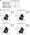
Induction of specific CTL by S M26–35 and SSL M26–35 in melanoma-derived TIL. Panel (A): CD8+ melanoma-derived TIL were stimulated in an LDA setting with decreasing amounts of S M26–35 or SSL M26–35 antigens. cytotoxic T-cells precursor frequencies are expressed as the number of specific effectors per 106 CD8+ cells. Panel (B): CD8+ melanoma-derived TIL stimulated in bulk cultures in the presence of the indicated antigen formulations and concentrations were stained with FITC-labelled anti-CD8 and PE-labelled M27–35 tetramers, as described in ‘Materials and methods’. Percentages of tetramer-positive CD8+ cells and the respective MFI are reported.

References

    1. Agrawal B, Krantz MJ, Reddish MA, Longenecker BM (1998) Rapid induction of primary CD4+ and CD8+ T cell responses against cancer-associated MUC-1 peptide epitopes. Int Immunol 157: 2089–2095 - PubMed
    1. Albo F, Cavazza A, Giardina B, Lippa S, Marini M, Roda LG, Spagnoli G (2003) Degradation of the immunogenic peptide gp100 280–288 by the monocyte-like U937 cell line. Peptides 24: 371–378 - PubMed
    1. Allen TM (1994) Long-circulating (sterically stabilized) liposomes for targeted drug delivery. Trends Pharmacol Sci 15: 215–220 - PubMed
    1. Alving CR, Koulchin V, Glenn GM, Rao M (1995) Liposomes as carriers of peptide antigens: induction of antibodies and cytotoxic T lymphocytes to conjugated and unconjugated peptides. Immunol Rev 145: 5–31 - PubMed
    1. Amoscato AA, Prenovitz DA, Lotze MT (1998) Rapid extracellular degradation of synthetic class I peptides by human dendritic cells. J Immunol 161: 4023–4032 - PubMed

Publication types