Review
. 2004 Jan 7:2:3.
doi: 10.1186/1477-7827-2-3.

Syndecans in tumor cell adhesion and signaling

Affiliations
Review

Syndecans in tumor cell adhesion and signaling

DeannaLee M Beauvais et al. Reprod Biol Endocrinol. .

Abstract

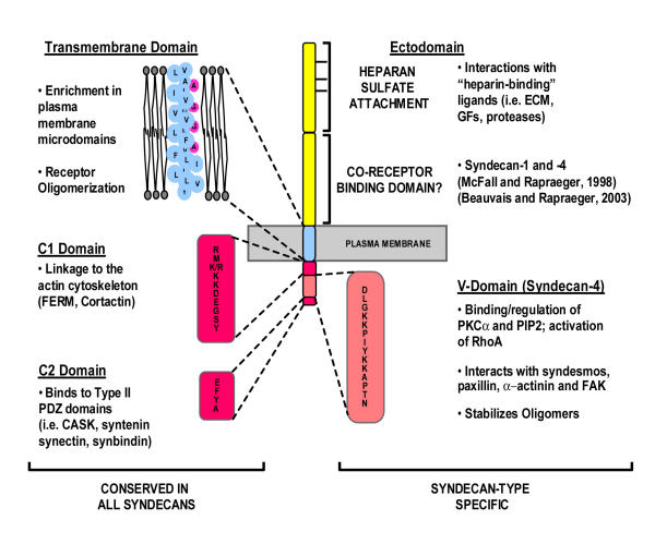
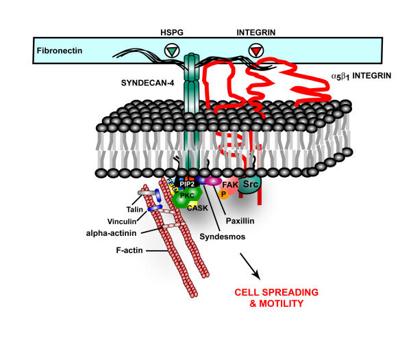
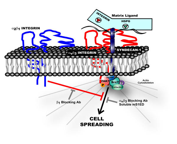
Anchorage of cells to "heparin"--binding domains that are prevalent in extracellular matrix (ECM) components is thought to occur primarily through the syndecans, a four-member family of transmembrane heparan sulfate proteoglycans that communicate environmental cues from the ECM to the cytoskeleton and the signaling apparatus of the cell. Known activities of the syndecans trace to their highly conserved cytoplasmic domains and to their heparan sulfate chains, which can serve to regulate the signaling of growth factors and morphogens. However, several emerging studies point to critical roles for the syndecans' extracellular protein domains in tumor cell behavior to include cell adhesion and invasion. Although the mechanisms of these activities remain largely unknown, one possibility involves "co-receptor" interactions with integrins that may regulate integrin function and the cell adhesion-signaling phenotype. Thus, alterations in syndecan expression, leading to either overexpression or loss of expression, both of which take place in tumor cells, may have dramatic effects on tumor cell invasion.


Figures

Figure 1
Figure 1
Syndecan Functional Domains. The syndecan core proteins display a high degree of conservation across species and across family members within their transmembrane and cytoplasmic domains, implying that some functional redundancy may exist among the family members. In contrast, the ectodomains of the syndecan core proteins are highly divergent both in size and sequence, indicating that this domain may confer separate and unique functions to each individual family member. Specific functions for the ectodomain of the syndecan core proteins are almost wholly unknown.
Figure 2
Figure 2
Syndecan-4 provides a mechanical link between the extracellular matrix and the actin cytoskeleton via binding interactions of its cytoplasmic tail. PIP2 binds to the syndecan-4 V-domain – an interaction that stabilizes syndecan-4 multimers against the inner leaflet of the plasma membrane and potentiates PKCα activity. The cytoplasmic tail also interacts with structural and signaling proteins such as CASK, FAK, syndesmos and paxillin. Interactions with these proteins may provide both a mechanical and signaling link to cell surface α5β1 integrins required for focal adhesion and stress fiber formation in cells adherent to fibronectin. Syndecan-4 may also link directly to the actin cytoskeleton through CASK, α-actinin and the FERM family of actin-binding proteins. Less is understood about the functional role of the V-domains of the other syndecan family members. However, the low degree of conservation within this domain amongst the four syndecans suggests distinct roles for the cytoplasmic domains of each of the syndecans in adhesion and signaling.
Figure 3
Figure 3
Syndecan-1 adhesion-mediated signaling in human mammary carcinomas. In response to syndecan-1 ligation, αvβ3 integrins and syndecan-1 form a signaling complex that regulates cytoskeletal reorganization. Cells initially fail to spread in response to syndecan-1 ligation because signaling downstream of this complex is negatively regulated by activated α2β1 integrins. Treatment of cells with a neutralizing β1 integrin antibody relieves this inhibitory signal allowing cells to initiate a spreading response. Intriguingly, signaling occurs in the absence of an integrin ligand suggesting that syndecan-1 participates in the allosteric activation of αvβ3 integrins. While activated αvβ3 integrins are required for signaling (i.e. spreading is blocked by αvβ3 integrin blocking antibody), molecular interactions of the syndecan-1 core protein are also required as signaling is blocked by soluble murine syndecan-1ectodomain (mS1ED). In our model, the syndecan physically associates with the αvβ3 integrin via an ectodomain interaction, although a functional coupling via an indirect mechanism has not been ruled out. Further, while our data suggest that the syndecan's transmembrane and cytoplasmic domains are not absolutely required for signaling, the possibility that these domains may have important regulatory roles cannot be excluded.

References

    1. Brooks PC, Montgomery AM, Rosenfeld M, Reisfeld RA, Hu T, Klier G, Cheresh DA. Integrin alpha v beta 3 antagonists promote tumor regression by inducing apoptosis of angiogenic blood vessels. Cell. 1994;79:1157–1164. - PubMed
    1. Domanico SZ, Pelletier AJ, Havran WL, Quaranta V. Integrin alpha 6A beta 1 induces CD81-dependent cell motility without engaging the extracellular matrix migration substrate. Mol Biol Cell. 1997;8:2253–2265. - PMC - PubMed
    1. Brassard DL, Maxwell E, Malkowski M, Nagabhushan TL, Kumar CC, Armstrong L. Integrin alpha(v)beta(3)-mediated activation of apoptosis. Exp Cell Res. 1999;251:33–45. - PubMed
    1. Kuzuya M, Satake S, Ramos MA, Kanda S, Koike T, Yoshino K, Ikeda S, Iguchi A. Induction of apoptotic cell death in vascular endothelial cells cultured in three-dimensional collagen lattice. Exp Cell Res. 1999;248:498–508. - PubMed
    1. Stupack DG, Puente XS, Boutsaboualoy S, Storgard CM, Cheresh DA. Apoptosis of adherent cells by recruitment of caspase-8 to unligated integrins. J Cell Biol. 2001;155:459–470. - PMC - PubMed

LinkOut - more resources