. 2004 Jan;70(1):346-55.
doi: 10.1128/AEM.70.1.346-355.2004.

Frequency, size, and localization of bacterial aggregates on bean leaf surfaces

Affiliations

Frequency, size, and localization of bacterial aggregates on bean leaf surfaces

J-M Monier et al. Appl Environ Microbiol. 2004 Jan.

Abstract

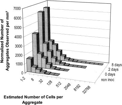
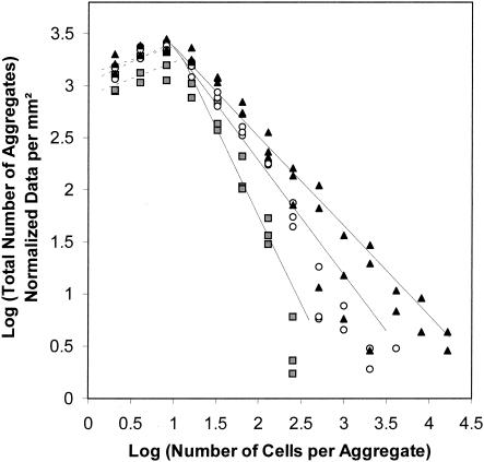
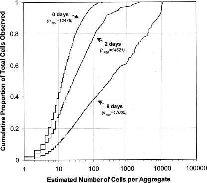
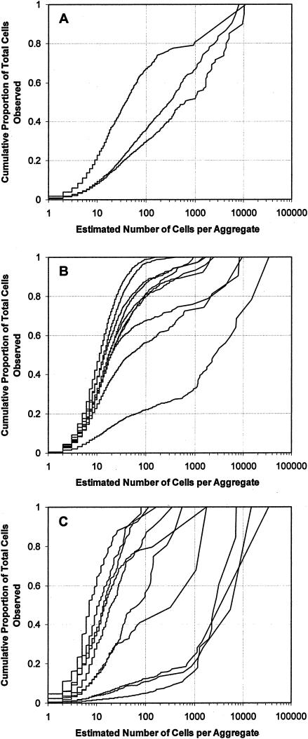
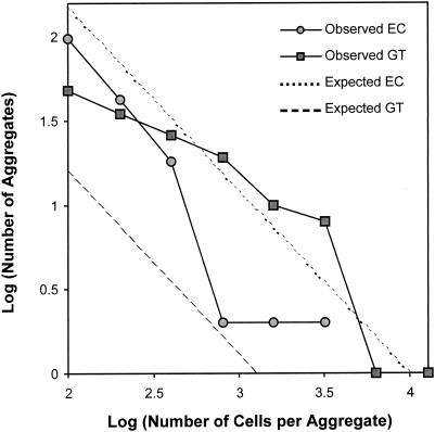
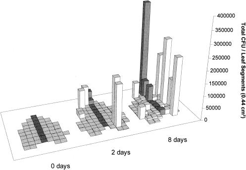
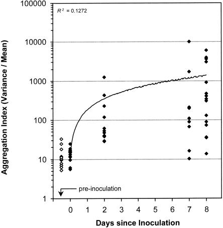
Using epifluorescence microscopy and image analysis, we have quantitatively described the frequency, size, and spatial distribution of bacterial aggregates on leaf surfaces of greenhouse-grown bean plants inoculated with the plant-pathogenic bacterium Pseudomonas syringae pv. syringae strain B728a. Bacterial cells were not randomly distributed on the leaf surface but occurred in a wide range of cluster sizes, ranging from single cells to over 10(4) cells per aggregate. The average cluster size increased through time, and aggregates were more numerous and larger when plants were maintained under conditions of high relative humidity levels than under dry conditions. The large majority of aggregates observed were small (less than 100 cells), and aggregate sizes exhibited a strong right-hand-skewed frequency distribution. While large aggregates are not frequent on a given leaf, they often accounted for the majority of cells present. We observed that up to 50% of cells present on a leaf were located in aggregates containing 10(3) cells or more. Aggregates were associated with several different anatomical features of the leaf surface but not with stomates. Aggregates were preferentially associated with glandular trichomes and veins. The biological and ecological significance of aggregate formation by epiphytic bacteria is discussed.


Figures

FIG. 1.
FIG. 1.
Frequency distribution of bacterial aggregates on leaf surfaces before inoculation and 0, 2, and 8 days following inoculation with P. syringae pv. syringae strain B728a. The total number of aggregates was determined for three leaves at each sampling time, and the data were combined. The characteristics of the total surface of the leaf observed at each sampling time were slightly different, and the total number of aggregates observed was normalized; values are expressed in square millimeters. The number of cells per aggregate was estimated from the surface area of each aggregate; it was assumed that each aggregate consisted of a dense monolayer of bacterial cells with an average surface area of 1.5 μm2.
FIG. 2.
FIG. 2.
Frequency distribution of aggregate sizes at 0 (gray-shaded squares), 2 (open circles), and 8 (filled triangles) days following inoculation with P. syringae pv. syringae strain B728a on bean leaf surfaces. Three leaves were observed at each sampling time. The TPL were used to compare aggregation patterns as a function of time. The cutoff sizes were 12, 19, and 10 cells per aggregate after 0, 2, and 8 days, respectively. The slope of the second TPL functions significantly (P < 0.001) decreased through time, reaching −1.67 (R2 = 0.88), −1.09 (R2 = 0.89), and −0.86 (R2 = 0.86) after 0, 2, and 8 days, respectively. No significant differences were observed between the slopes of the first power functions corresponding to the smaller aggregates.
FIG. 3.
FIG. 3.
Cumulative proportion of the total number of cells observed as a function of the total number of cells per aggregate. Each curve represents the combined data of three individual leaves observed 0, 2, and 8 days following inoculation with P. syringae pv. syringae strain B728a.
FIG. 4.
FIG. 4.
Cumulative proportion of the total number of cells observed as a function of the total number of cells per aggregate when considered over different spatial scales. Each curve corresponds to the results for an individual leaf (A), an individual leaf segment cut randomly within the same leaf (B), or an individual field of view randomly selected within the same leaf segment (C) observed 7 days following inoculation with P. syringae pv. syringae strain B728a.
FIG. 5.
FIG. 5.
Observed and expected frequency distributions of aggregate sizes associated with glandular trichomes (GT) and nondifferentiated epidermal cells (EC) 8 days following inoculation with P. syringae pv. syringae strain B728a on bean leaf surfaces. Aggregates associated with glandular trichomes were more numerous (P < 0.001) and significantly larger (P < 0.001) than expected. Aggregates associated with nondifferentiated cells were significantly smaller (P < 0.005) and less numerous (P < 0.001) than expected. The same results were observed 2 days after inoculation (data not shown).
FIG. 6.
FIG. 6.
Culturable population sizes of P. syringae pv. syringae strain B728a among different leaf segments cut from an individual bean leaf, 0, 2, and 8 days after inoculation. Bacteria were removed from individual 44-mm2 segments at each harvest time. The variance/mean ratios for populations on different segments collected 0, 2, and 8 days after inoculation with P. syringae were 971, 104,434, and 157,812, respectively.
FIG. 7.
FIG. 7.
Aggregation index (variance/mean) ratios within leaf segments as determined by image analysis as a function of time on noninoculated plants (open diamonds) and following inoculation with P. syringae pv. syringae strain B728a (filled diamonds). Each symbol represents an individual leaf segment.

References

    1. Andersen, G. L., O. Menkissoglou, and S. E. Lindow. 1991. Occurrence and properties of copper-tolerant strains of Pseudomonas syringae isolated from fruit trees in California [USA]. Phytopathology 81:648-656.
    1. Ascensao, L., and M. S. Pais. 1998. The leaf capitate trichomes of Leonotis leonurus: histochemistry, ultrastructure and secretion. Ann. Bot. 81:263-271.
    1. Bailey, M. J., A. K. Lilley, and J. P. Diaper. 1996. Gene transfer between microorganisms in the phyllosphere, p 103-123. In C. E. Morris, P. C. Nicot, and C. Nguyen-The (ed.), Aerial plant surface microbiology. Plenum Press, New York, N.Y.
    1. Barber, C. E., J. L. Tang, J. X. Feng, M. Q. Pan, T. J. G. Wilson, H. Slater, J. M. Dow, P. Williams, and M. J. Daniels. 1997. A novel regulatory system required for pathogenicity of Xanthomonas campestris is mediated by a small diffusible signal molecule. Mol. Microbiol. 24:555-566. - PubMed
    1. Beattie, G. A., and S. E. Lindow. 1994. Comparison of the behavior of epiphytic fitness mutants of Pseudomonas syringae under controlled and field conditions. Appl. Environ. Microbiol. 60:3799-3808. - PMC - PubMed

Publication types

MeSH terms