. 2004 Jan 20;101(3):847-52.
doi: 10.1073/pnas.0304375101. Epub 2004 Jan 7.

Mislocalization to the nuclear envelope: an effect of the dystonia-causing torsinA mutation

Affiliations

Mislocalization to the nuclear envelope: an effect of the dystonia-causing torsinA mutation

Rose E Goodchild et al. Proc Natl Acad Sci U S A. .

Abstract

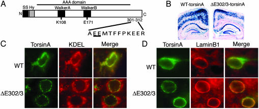
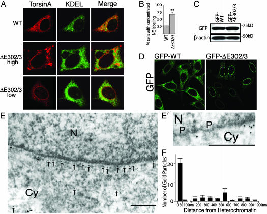
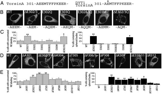
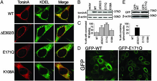
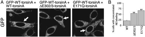
Primary dystonia is a disease characterized by involuntary twisting movements caused by CNS dysfunction without underlying histopathology. DYT1 dystonia is a form of primary dystonia caused by an in-frame GAG deletion (DeltaE302/3) in the TOR1A gene that encodes the endoplasmic reticulum luminal protein torsinA. We show that torsinA is also present in the nuclear envelope (NE), where it appears to interact with substrate, and that the DeltaE302/3 mutation causes a striking redistribution of torsinA from the endoplasmic reticulum to the NE. In addition, DeltaE302/3-torsinA recruits WT torsinA to the NE, potentially providing insight into an understanding of the dominant inheritance of the disease. DYT1 dystonia appears to be a previously uncharacterized NE disease and the first, to our knowledge, to selectively affect CNS function.


Figures

Fig. 1.
Fig. 1.
ΔE302/3-torsinA is abnormally localized in the neuronal NE. (A) Schematic representation of the torsinA protein. The primary sequence surrounding the DYT1 mutation is shown with the site of the glutamic acid deletion underlined. SS, signal sequence; Hy, hydrophobic domain; K108 and E171, site of key Walker box residues mutated for functional studies of torsinA. (B) β-Galactosidase histochemistry of brain sections from WT- and ΔE302/3-torsinA-expressing transgenic mice. (C and D) Frontal cortex sections from transgenic mice expressing either WT-torsinA or ΔE302/3-torsinA and double stained with antibodies against torsinA and KDEL (C) or laminB1 (D). No torsinA immunolabeling was observed in nonbitransgenic littermates (data not shown).
Fig. 2.
Fig. 2.
ΔE302/3-torsinA is concentrated in the NE in vitro.(A) Immunofluorescent labeling of BHK21 cells transiently transfected with WT- or ΔE302/3-torsinA. (Middle and Bottom) NE concentration of ΔE302/3-torsinA in both high and low ΔE302/3-torsinA-expressing cells. Only high ΔE302/3-torsinA-expressing cells displayed spheroid body accumulation of torsinA. No differences were observed between high and low WT-torsinA-expressing cells. (B) Percentage of transfected cells with concentrated NE torsinA labeling. Each cDNA transfection was performed three times; error bars represent SEM. **, Significant (P < 0.01) difference from WT. (C) SDS/PAGE of Triton X-100-extracted protein illustrating that GFP-tagged torsinA proteins were expressed at similar levels. (D) Direct GFP fluorescence from BHK cells stably expressing GFP-tagged WT-torsinA or ΔE302/3-torsinA. (E and E′) ImmunoGold electron microscopic localization of GFP in GFP-ΔE302/3-torsinA cell line. Gold particles are highlighted with arrows. N, nucleus; Cy, cytoplasm; P, nuclear pore complex. (Scale bars, 200 nm.) (F) Distance of gold particles from electron dense heterochromatin was measured in images from four randomly selected cells. Error bars represent SEM.
Fig. 3.
Fig. 3.
Multiple single amino acid deletions in torsinA mimic ΔE302/3-torsinA mislocalization to the NE. (A) Primary amino acid sequence of the C-terminal region surrounding the DYT1 mutation. (B) Immunofluorescent torsinA labeling of transiently transfected BHK21 cells. (C) Percentage of transfected cells with concentrated NE torsinA labeling or spheroid bodies. (D) Immunofluorescent torsinA labeling of BHK21 cells transiently transfected with versions of torsinA containing single amino acid deletions. (E) Percentage of transfected cells with concentrated NE torsinA labeling or spheroid bodies. All cDNA transfections were performed three times; error bars represent SEM. * and **, Significant (P < 0.05 and P < 0.01, respectively) difference from WT.
Fig. 4.
Fig. 4.
ATP-bound torsinA is selectively localized at the NE. (A) Immunofluorescent labeling of transiently transfected BHK21 cells. (B) SDS/PAGE of Triton X-100-extracted protein shows that the torsinA constructs were expressed at similar levels. (C) Percentage of transfected cells with concentrated NE torsinA labeling or spheroid bodies. Each cDNA transfection was performed three times; error bars represent SEM. **, Significant (P < 0.01) difference from WT. (D) Direct GFP fluorescence from BHK21 cells stably expressing GFP-tagged WT- or E171Q-torsinA. (E) SDS/PAGE of Triton X-100-extracted protein demonstrating that GFP-tagged torsinA proteins were expressed at similar levels.
Fig. 5.
Fig. 5.
ΔE302/3-torsinA recruits WT-torsinA to the NE. (A) Distribution of GFP immunoreactivity in the GFP-WT-torsinA cell line transfected with WT-, ΔE302/3-, or E171Q-torsinA. Arrows indicate transfected cells (identified by β-galactosidase expression). (B) Quantification of the percentage of transfected cells with clearly enhanced NE GFP immunolabeling (compared to untransfected GFP-WT-torsinA cells). Quantification was performed by a blinded observer assessing the NE concentration of GFP labeling in 100 β-galactosidase-positive cells. Each cDNA transfection was performed three times; error bars represent SEM. **, Significant (P < 0.01) difference from WT transfected GFP-WT-torsinA cells.
Fig. 6.
Fig. 6.
TorsinA abnormally localizes to the NE in tissue from DYT1 patients. (A) Immunofluorescent torsinA labeling of control and DYT1 patient fibroblasts. (B) SDS/PAGE of Triton X-100-extracted protein probed with anti-torsinA and anti-β-actin antibodies.

Comment in

References

    1. Fahn, S., Marsden, C. D. & Calne, D. B. (1987) in Movement Disorders 2, eds. Marsden, C. D. & Fahn, S. (Butterworths, London), pp. 332–58.
    1. Berardelli, A., Rothwell, J. C., Hallett, M., Thompson, P. D., Manfredi, M. & Marsden, C. D. (1998) Brain 121, 1195–1212. - PubMed
    1. Ozelius, L. J., Hewett, J. W., Page, C. E., Bressman, S. B., Kramer, P. L., Shalish, C., de Leon, D., Brin, M. F., Raymond, D., Corey, D. P., et al. (1997) Nat. Genet. 17, 40–48. - PubMed
    1. Neuwald, A. F., Aravind, L., Spouge, J. L. & Koonin, E. V. (1999) Genome Res. 9, 27–43. - PubMed
    1. Ogura, T. & Wilkinson, A. J. (2001) Genes Cells 6, 575–597. - PubMed

Publication types

MeSH terms