. 2004 Jan 14;23(1):169-79.
doi: 10.1038/sj.emboj.7600030. Epub 2004 Jan 8.

Cytoprotection by pre-emptive conditional phosphorylation of translation initiation factor 2

Affiliations

Cytoprotection by pre-emptive conditional phosphorylation of translation initiation factor 2

Phoebe D Lu et al. EMBO J. .

Abstract

Transient phosphorylation of the alpha-subunit of translation initiation factor 2 (eIF2alpha) represses translation and activates select gene expression under diverse stressful conditions. Defects in the eIF2alpha phosphorylation-dependent integrated stress response impair resistance to accumulation of malfolded proteins in the endoplasmic reticulum (ER stress), to oxidative stress and to nutrient deprivations. To study the hypothesized protective role of eIF2alpha phosphorylation in isolation of parallel stress signaling pathways, we fused the kinase domain of pancreatic endoplasmic reticulum kinase (PERK), an ER stress-inducible eIF2alpha kinase that is normally activated by dimerization, to a protein module that binds a small dimerizer molecule. The activity of this artificial eIF2alpha kinase, Fv2E-PERK, is subordinate to the dimerizer and is uncoupled from upstream stress signaling. Fv2E-PERK activation enhanced the expression of numerous stress-induced genes and protected cells from the lethal effects of oxidants, peroxynitrite donors and ER stress. Our findings indicate that eIF2alpha phosphorylation can initiate signaling in a cytoprotective gene expression pathway independently of other parallel stress-induced signals and that activation of this pathway can single-handedly promote a stress-resistant preconditioned state.


Figures

Figure 1
Figure 1
Stresses that promote eIF2α phosphorylation protect HT22 cells from glutamate-induced cell death. (A) Survival of HT22 cells pretreated for 1 h with thapsigargin or tunicamycin followed by 5 h of recovery and subsequent challenge with 5 mM glutamate (closed squares) or 10 mM glutamate (open squares) for 24 h. Survival is expressed as a percentage of MTT reduced by glutamate-treated cells in each pretreatment group compared with MTT reduction by cells subjected to the same pretreatment but not exposed to glutamate. Values shown are the mean and SEM from a representative experiment performed in triplicate and reproduced twice. (B) Immunoblots of cytoplasmic extracts incubated with antisera specific for phosphorylated eIF2α (eIF2α-P), total eIF2α and GADD34 and of nuclear extracts incubated with antisera to ATF4 and CHOP from untreated HT22 cells, or cells treated with thapsigargin (Tg, 0.5 μM) or tunicamycin (Tm, 0.3 μg/ml) for 0.5 or 1 h and after 5 h of recovery (R).
Figure 2
Figure 2
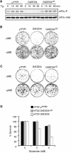
Impaired eIF2α phosphorylation sensitizes HT22 cells to oxidative glutamate toxicity. (A) Immunoblot of phosphorylated eIF2α (eIF2α-P) and total eIF2α from ψempty, GADD34 or GADD34ΔC cells treated with 400 nM thapsigargin (Tg) for the indicated time. (B) Photomicrograph of crystal-violet-stained HT22 cells transduced with retroviruses expressing an activated form of an eIF2α phosphatase (GADD34), an inactive mutant (GADD34ΔC) or the empty vector (ψempty). Following transduction, cells were plated at clonal density and cultured in regular media or media supplemented with the reducing compound βME for 6 days prior to staining. (C) Photomicrograph of crystal-violet-stained HT22 cells selected for stable expression of GADD34, ψempty or GADD34ΔC and plated at clonal density regular in media or media containing βME. (D) Survival of HT22 GADD34, ψempty or GADD34ΔC cells following 24 h of exposure to glutamate in media lacking βME. 100% survival is defined as the MTT reduction by cells of each line that had not been exposed to glutamate. The graph shown represents a typical experiment performed in duplicate and reproduced twice.
Figure 3
Figure 3
Uncoupling the eIF2α kinase activity of PERK from its activation by ER stress. (A) Schematic representation of the Fv2E-PERK fusion protein. The modified FKBP domains (Fv) and the transmembrane domain of PERK (TM) are indicated. (B) Immunoblot of Fv2E-PERK, phosphorylated (eIF2α-P) and total eIF2α, and the downstream targets GADD34 and CHOP in lysates of Fv2E-PERK-expressing HT22 cells and parental HT22 cells following treatment with 1 nM of the AP20187 dimerizer or thapsigargin (Tg). The position of the activated and phosphorylated (Fv2E-PERKP) and the inactive hypophosphorylated Fv2E-PERK (Fv2E-PERK0) are indicated. (C) Autoradiogram of 35S-methionine incorporation into newly synthesized proteins in Fv2E-PERK-expressing HT22 cells that had been left untreated (UT) or had been treated with 1 nM AP20187 for 1 h or 400 nM thapsigargin (Tg) for 0.5 h. (D) Immunoblot of Fv2E-PERK, phosphorylated (eIF2α-P) and total eIF2α, and the downstream targets GADD34 and ATF4 in lysates of Fv2E-PERK-expressing MEFs with wild-type (S/S) and mutant (A/A) eIF2α genotypes, following treatment with 2 nM of the AP20187 dimerizer. (E) Microarray analysis of the expression level of ISR target genes in Fv2E-PERK MEFs with wild-type (S/S) and mutant (A/A) eIF2α genotypes. The 375 genes induced at least two-fold in both AP20187-treated Fv2E-PERK MEFs (at either 4 or 8 h) and tunicamycin-treated wild-type MEFs or in tunicamycin-treated MEFs alone are shown. The expression levels of the genes are shaded, with white indicating low levels of expression and black indicating high levels of expression. The genes are clustered to reveal a group of PERK-dependent tunicamycin-induced genes that are also activated by Fv2E-PERK (Group A) and a group of PERK-dependent tunicamycin-induced genes that are not induced by Fv2E-PERK activation (Group B).
Figure 4
Figure 4
Activation of Fv2E-PERK enhances survival of glutamate-exposed HT22 cells. (A) Immunoblot of Fv2E-PERK, phosphorylated eIF2α (eIF2α-P), total eIF2α and the downstream target GADD34 in untreated cells (UT) and at various points of recovery following a 15-min exposure to 1 or 2 nM AP20187. The position of the activated Fv2E-PERKP and inactive Fv2E-PERK0 proteins is indicated. (B) 35S-methionine incorporation into newly synthesized proteins at various time points of recovery from translational inhibition by treatment for 15 min with 1 nM AP20187 (circles) or 2 nM AP20187 (squares). Translation is expressed as the percentage of 35S-methionine incorporation into untreated cells, which is arbitrarily set at 100%. (C) Survival of parental HT22 cells or cells expressing Fv2E-PERK that had been treated with 1 nM, or 2 nM AP20187 or ethanol carrier for 15 min followed by 5 or 12 h of recovery and subsequently exposed to the indicated concentration of glutamate for 24 h. 100% survival is defined as the MTT reduction in cells that had not been exposed to glutamate in each AP20187 treatment group. The means±SEM of a representative experiment performed in duplicate and repeated four times are shown. (D) Photomicrographs of crystal-violet-stained parental HT22 cells or cells expressing Fv2E-PERK treated with 1 or 2 nM AP20187 or ethanol carrier for 1 h followed by 5 h of recovery and subsequently exposed to glutamate for 24 h. The cells were switched to regular media for 6 days and then stained with crystal violet.
Figure 5
Figure 5
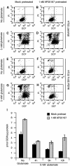
AP20187-mediated activation of Fv2E-PERK reduces the levels of glutamate-induced endogenous peroxides and rescues glutamate-induced glutathione depletion. Dual-channel FACscans of dichlorofluorescein fluorescence (X-axis) and propidium iodide fluorescence (Y-axis) of parental HT22 cells (AD) or Fv2E-PERK cells (EH) that were either mock pretreated (A, C, E, G) or pretreated with 1 nM AP20187 for 1 h followed by a 5 h recovery period (B, D, F, H) before exposure to 5 mM glutamate for 10 h (C, D, G, H) are shown. The fraction of cells in each quadrant of the two-dimensional FACscan is displayed in the table below. [Table: see text] (I) Glutathione levels in HT22 cells expressing Fv2E-PERK either mock pretreated or pretreated with 1 nM AP20187 followed by a 5 h recovery period. Glutathione levels were measured at various points after exposure to 5 mM glutamate or at 10 h postrecovery in the absence of glutamate. The means±SEM (n=2) are shown.
Figure 6
Figure 6
AP20187-induced activation of Fv2E-PERK in MEFs is protective against ER stress and nitrosylation stress. (A) Immunoblot of Fv2E-PERK from Wildtype (Perk+/+) and Perk−/− cells selected for stable expression of Fv2E-PERK and treated with 1 nM AP20187 for the indicated periods of time. The positions of the activated and phosphorylated (Fv2E-PERKP) and the inactive hypophosphorylated Fv2E-PERK (Fv2E-PERK0) are indicated. (B) Immunoblot analysis of immunoprecipitated endogenous PERK, phosphorylated eIF2α (eIF2α-P), total eIF2α, ATF4, and CHOP from Perk+/+ and Perk−/− cells expressing Fv2E-PERK and treated with 1 nM AP20187 or 400 nM thapsigargin (Tg). The positions of the activated and phosphorylated (endogenous PERKP) and the inactive hypophosphorylated PERK (endogenous PERK0) are indicated. (C) Photomicrographs of crystal-violet-stained cells with wild-type (S/S) or mutant (A/A) eIF2α genotypes expressing Fv2E-PERK treated with AP20187 or ethanol carrier prior to exposure to tunicamycin (Tm) for 16 h as described in Methods. The media were replaced after tunicamycin exposure and the cells were stained with crystal violet 6 days later. (D) Survival of cells with wild-type (S/S) or mutant (A/A) eIF2α genotypes expressing Fv2E-PERK that had been pretreated with 1 nM AP20187 or ethanol carrier and exposed to SIN-1 at the indicated concentrations. 100% survival is defined as the MTT reduction in cells that had not been exposed to SIN-1. The means±SEM of a representative experiment performed in triplicate and repeated twice are shown.

References

    1. Anderson ME 1985. Determination of glutathione and glutathione disulfide in biological samples. Methods Enzymol 113: 548–555 - PubMed
    1. Bertolotti A, Zhang Y, Hendershot L, Harding H, Ron D 2000. Dynamic interaction of BiP and the ER stress transducers in the unfolded protein response. Nat Cell Biol 2: 326–332 - PubMed
    1. Brostrom CO, Brostrom MA 1998. Regulation of translational initiation during cellular responses to stress. Prog Nucleic Acid Res Mol Biol 58: 79–125 - PubMed
    1. Chen J 2000. Heme-regulated eIF2α kinase. In Translational Control of Gene Expression, Sonenberg N, Hershey JWB, and Mathews MB (eds) pp 529–546. Cold Spring Harbor: CSHL Press
    1. Davis JB, Maher P 1994. Protein kinase C activation inhibits glutamate-induced cytotoxicity in a neuronal cell line. Brain Res 652: 169–173 - PubMed

Publication types

MeSH terms