

Macular pigment Raman detector for clinical applications
- PMID: 14715066
- PMCID: PMC3086335
- DOI: 10.1117/1.1627776
Macular pigment Raman detector for clinical applications
Abstract
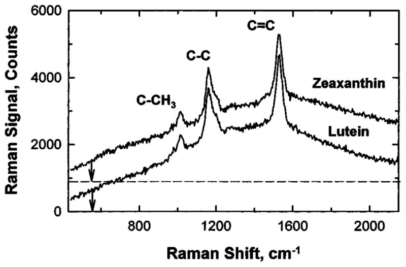
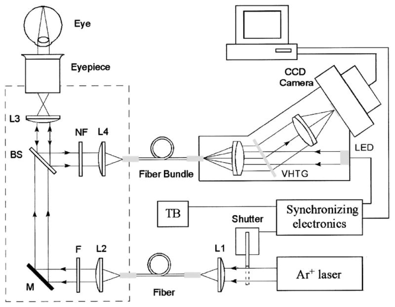
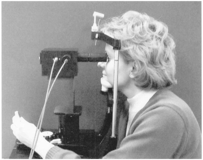
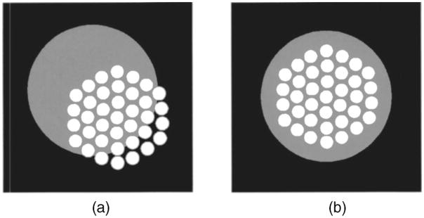
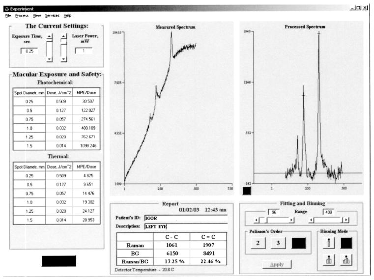
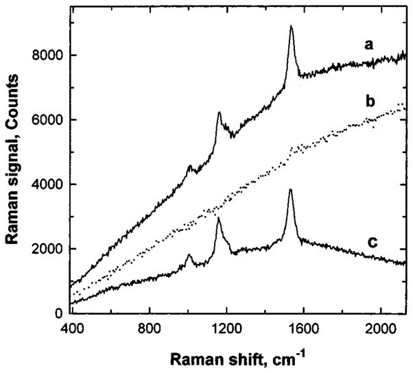
Clinical studies of carotenoid macular pigments (MP) have been limited by the lack of noninvasive, objective instruments. We introduce a novel noninvasive optical instrument, an MP Raman detector, for assessment of the carotenoid status of the human retina in vivo. The instrument uses resonant excitation of carotenoid molecules in the visible wavelength range, and quantitatively measures the highly specific Raman signals that originate from the single- and double-bond stretch vibrations of the pi-conjugated carotenoid molecule's carbon backbone. The instrument is a robust, compact device and suitable for routine measurements of MP concentrations in a clinical setting. We characterized and tested the instrument in clinical studies of human subjects to validate its function and to begin to establish its role as a possible screening test for macular pathologies. We also show that the MP Raman spectroscopy technology has potential as a novel, highly specific method for rapid screening of carotenoid antioxidant levels in large populations at risk for vision loss from age-related macular degeneration, the leading cause of blindness of the elderly in the developed world.
(c) 2004 Society of Photo-Optical Instrumentation Engineers.
Figures
Similar articles
-
Resonance Raman detection of carotenoid antioxidants in living human tissue.J Biomed Opt. 2005 Nov-Dec;10(6):064028. doi: 10.1117/1.2139974. J Biomed Opt. 2005. PMID: 16409093 Free PMC article. Review.
-
Simple Raman instrument for in vivo detection of macular pigments.Appl Spectrosc. 2005 Jul;59(7):861-7. doi: 10.1366/0003702054411616. Appl Spectrosc. 2005. PMID: 16053555 Free PMC article.
-
In vivo resonant Raman measurement of macular carotenoid pigments in the young and the aging human retina.J Opt Soc Am A Opt Image Sci Vis. 2002 Jun;19(6):1172-86. doi: 10.1364/josaa.19.001172. J Opt Soc Am A Opt Image Sci Vis. 2002. PMID: 12049355
-
Noninvasive detection of macular pigments in the human eye.J Biomed Opt. 2004 Jan-Feb;9(1):75-85. doi: 10.1117/1.1628240. J Biomed Opt. 2004. PMID: 14715058 Review.
-
Raman detection of macular carotenoid pigments in intact human retina.Invest Ophthalmol Vis Sci. 1998 Oct;39(11):2003-11. Invest Ophthalmol Vis Sci. 1998. PMID: 9761278
Cited by
-
Brighter is better: bill fluorescence increases social attraction in a colonial seabird and reveals a potential link with foraging.Behav Ecol Sociobiol. 2021 Oct;75(10):144. doi: 10.1007/s00265-021-03087-0. Epub 2021 Sep 25. Behav Ecol Sociobiol. 2021. PMID: 34840402 Free PMC article.
-
The value of measurement of macular carotenoid pigment optical densities and distributions in age-related macular degeneration and other retinal disorders.Vision Res. 2010 Mar 31;50(7):716-28. doi: 10.1016/j.visres.2009.10.014. Epub 2009 Oct 23. Vision Res. 2010. PMID: 19854211 Free PMC article. Review.
-
Effect of age and other factors on macular pigment optical density measured with resonance Raman spectroscopy.Graefes Arch Clin Exp Ophthalmol. 2014 Aug;252(8):1221-8. doi: 10.1007/s00417-014-2574-x. Epub 2014 Mar 11. Graefes Arch Clin Exp Ophthalmol. 2014. PMID: 24614948
-
Changes in Macular Pigment Optical Density and Serum Lutein Concentration in Japanese Subjects Taking Two Different Lutein Supplements.PLoS One. 2015 Oct 9;10(10):e0139257. doi: 10.1371/journal.pone.0139257. eCollection 2015. PLoS One. 2015. PMID: 26451726 Free PMC article. Clinical Trial.
-
Resonance Raman detection of carotenoid antioxidants in living human tissue.J Biomed Opt. 2005 Nov-Dec;10(6):064028. doi: 10.1117/1.2139974. J Biomed Opt. 2005. PMID: 16409093 Free PMC article. Review.
References
-
- Schalch W, Dayhaw-Barker P, Barker FM. The carotenoids of the human retina. In: Taylor A, editor. Nutritional and Environmental Influences on the Eye. CRC Press; Boca Raton, FL: 1999. pp. 215–250.
-
- Handelman GJ, Snodderly DM, Adler AJ, Russett MD, Dratz EA. Measurement of carotenoids in human and monkey retinas. Methods Enzymol. 1992;213:220–230. - PubMed
-
- Beatty S, Koh HH, Henson D, Boulton M. The role of oxidative stress in the pathogenesis of age-related macular degeneration. Surv Ophthalmol. 2000;45:115–134. - PubMed
-
- Reading VM, Weale RA. Macular pigment and chromatic aberration. J Am Optom Assoc. 1974;64:231–234. - PubMed
-
- Beatty S, Murray IJ, Henson DB, Carden D, Koh HH, Boulton ME. Macular pigment and risk for age-related macular degeneration in subjects from a northern European population. Invest Ophthalmol Visual Sci. 2001;42:439–446. - PubMed
MeSH terms
Substances
Grants and funding
LinkOut - more resources
Full Text Sources
Other Literature Sources
Medical
Research Materials
Miscellaneous
