

Novel non-viral method for transfection of primary leukemia cells and cell lines
- PMID: 14715084
- PMCID: PMC331421
- DOI: 10.1186/1479-0556-2-1
Novel non-viral method for transfection of primary leukemia cells and cell lines
Abstract
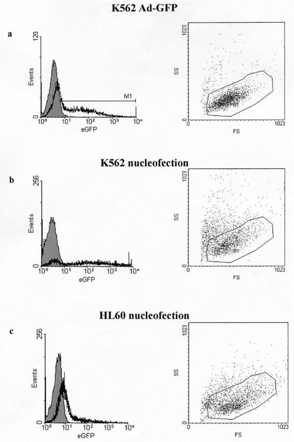
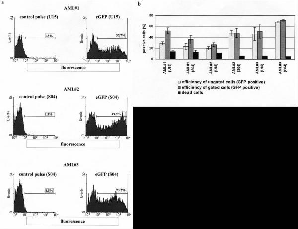
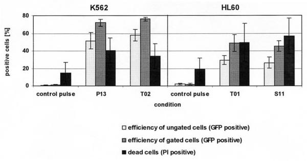
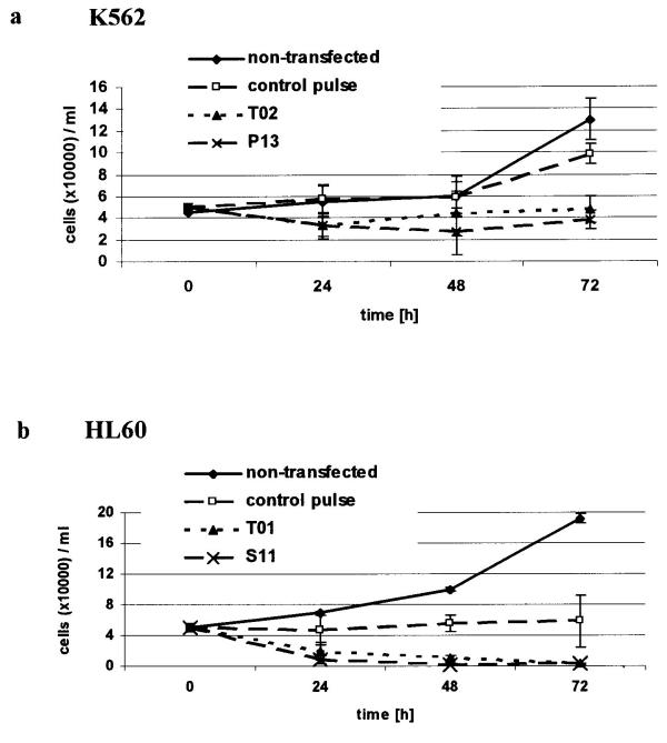
BACKGROUND: Tumor cells such as leukemia and lymphoma cells are possible targets for gene therapy. However, previously leukemia and lymphoma cells have been demonstrated to be resistant to most of non-viral gene transfer methods. METHODS: The aim of this study was to analyze various methods for transfection of primary leukemia cells and leukemia cell lines and to improve the efficiency of gene delivery. Here, we evaluated a novel electroporation based technique called nucleofection. This novel technique uses a combination of special electrical parameters and specific solutions to deliver the DNA directly to the cell nucleus under mild conditions. RESULTS: Using this technique for gene transfer up to 75% of primary cells derived from three acute myeloid leukemia (AML) patients and K562 cells were transfected with the green flourescent protein (GFP) reporter gene with low cytotoxicity. In addition, 49(+/- 9.7%) of HL60 leukemia cells showed expression of GFP. CONCLUSION: The non-viral transfection method described here may have an impact on the use of primary leukemia cells and leukemia cell lines in cancer gene therapy.
Figures
Similar articles
-
Spatial and Temporal Control of Cavitation Allows High In Vitro Transfection Efficiency in the Absence of Transfection Reagents or Contrast Agents.PLoS One. 2015 Aug 14;10(8):e0134247. doi: 10.1371/journal.pone.0134247. eCollection 2015. PLoS One. 2015. PMID: 26274324 Free PMC article.
-
Gene transfer to primary acute myeloid leukaemia blasts and myeloid leukaemia cell lines.Cytokines Cell Mol Ther. 2000 Sep;6(3):127-34. doi: 10.1080/mccm.6.3.127.134. Cytokines Cell Mol Ther. 2000. PMID: 11140881
-
Efficient gene expression in megakaryocytic cell line using nucleofection.Int J Pharm. 2007 Jun 29;338(1-2):157-64. doi: 10.1016/j.ijpharm.2007.01.042. Epub 2007 Feb 2. Int J Pharm. 2007. PMID: 17331684
-
Nucleofection is a highly effective gene transfer technique for human melanoma cell lines.Exp Dermatol. 2008 May;17(5):405-11. doi: 10.1111/j.1600-0625.2007.00687.x. Epub 2008 Feb 27. Exp Dermatol. 2008. PMID: 18312380
-
Review of alterations of the cyclin-dependent kinase inhibitor INK4 family genes p15, p16, p18 and p19 in human leukemia-lymphoma cells.Leukemia. 1998 Jun;12(6):845-59. doi: 10.1038/sj.leu.2401043. Leukemia. 1998. PMID: 9639410 Review.
Cited by
-
Targeted nanoparticles enhanced flow electroporation of antisense oligonucleotides in leukemia cells.Biosens Bioelectron. 2010 Oct 15;26(2):778-83. doi: 10.1016/j.bios.2010.06.025. Epub 2010 Jul 1. Biosens Bioelectron. 2010. PMID: 20630739 Free PMC article.
-
Phosphate-buffered saline-based nucleofection of primary endothelial cells.Anal Biochem. 2009 Mar 15;386(2):251-5. doi: 10.1016/j.ab.2008.12.021. Epub 2008 Dec 25. Anal Biochem. 2009. PMID: 19150324 Free PMC article.
-
Cell type differences in activity of the Streptomyces bacteriophage phiC31 integrase.Nucleic Acids Res. 2008 Oct;36(17):5462-71. doi: 10.1093/nar/gkn532. Epub 2008 Aug 21. Nucleic Acids Res. 2008. PMID: 18718925 Free PMC article.
-
Improvement of K562 Cell Line Transduction by FBS Mediated Attachment to the Cell Culture Plate.Biomed Res Int. 2019 Mar 27;2019:9540702. doi: 10.1155/2019/9540702. eCollection 2019. Biomed Res Int. 2019. PMID: 31032368 Free PMC article.
-
Clinical evaluation of cellular immunotherapy in acute myeloid leukaemia.Cancer Immunol Immunother. 2011 Jun;60(6):757-69. doi: 10.1007/s00262-011-1022-6. Epub 2011 Apr 26. Cancer Immunol Immunother. 2011. PMID: 21519825 Free PMC article. Review.
References
-
- Adams SW, Emerson SG. Gene therapy for leukemia and lymphoma. Hematol Oncol Clin North Am. 1998;12:631–648. - PubMed
-
- Einhorn S, Strander H. Interferon treatment of human malignancies – a short review. Med Oncol Tumor Pharmacother. 1993;10:25–29. - PubMed
-
- Finke S, Trojaneck B, Lefterova P, Csipai M, Wagner E, Neubauer A, Huhn D, Wittig B, Schmidt-Wolf IGH. Increase of proliferation rate and enhancement of antitumor cytotoxicity of human CD3+CD56+ immunologic effector cells by receptor-mediated transfection with the interleukin-7 gene. Gene Therapy. 1998;5:31–39. doi: 10.1038/sj.gt.3300560. - DOI - PubMed
-
- Zheng Z, Takahashi M, Aoki S, Toba K, Liu A, Osman Y, Takahashi H, Tsukada N, Suzuki N, Nikkuni K, Furukawa T, Koike T, Aizawa Y. Expression patterns of costimulatory molecules on cells derived from human hematological malignancies. J Exp Clin Cancer Res. 1998;17:251–258. - PubMed
LinkOut - more resources
Full Text Sources
