. 2004 Jan;11(1):152-60.
doi: 10.1128/cdli.11.1.152-160.2004.

Systemic and mucosal responses to oral administration of excretory and secretory antigens from Giardia intestinalis

Affiliations

Systemic and mucosal responses to oral administration of excretory and secretory antigens from Giardia intestinalis

Juan Carlos Jiménez et al. Clin Diagn Lab Immunol. 2004 Jan.

Abstract

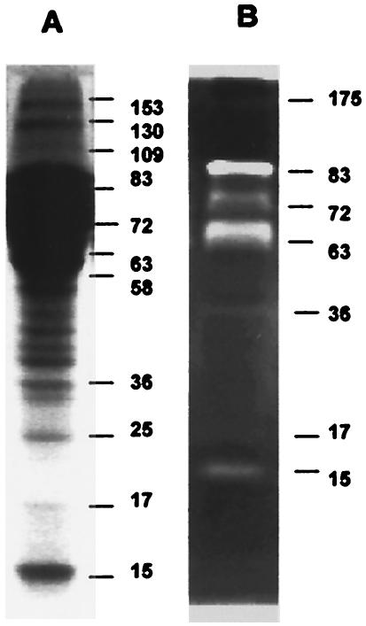
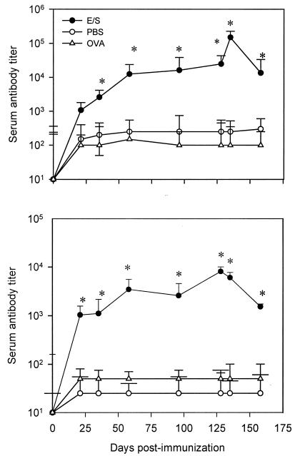
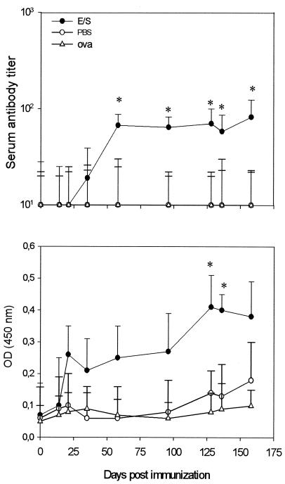
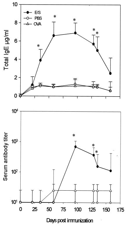
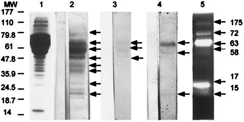
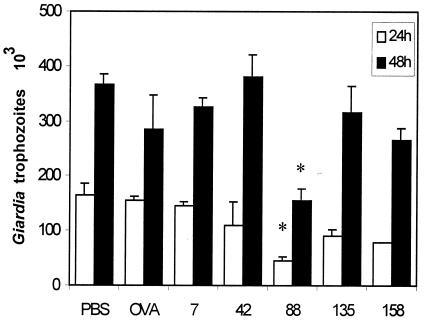
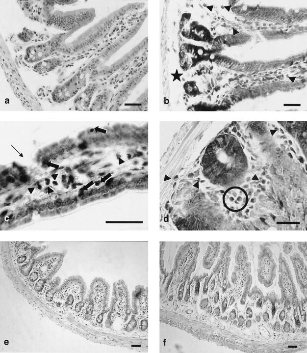
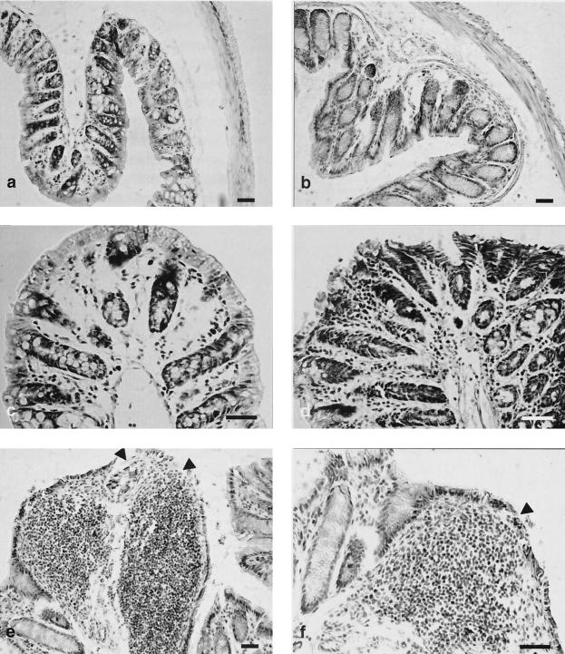
Giardia, a flagellated protozoan that infects the upper small intestine of its vertebrate host, is the most common parasitic protist responsible for diarrhea worldwide. Molecules released by the parasite, particularly excretory and secretory antigens, seemed to be associated with pathogenesis as well as with the expression of Giardia virulence. In the present work, we examined the effect of oral administration of Giardia intestinalis excretory and secretory antigens on systemic and local antibody response as well as on mucosal injuries in BALB/c mice. Significant titers of serum-specific immunoglobulin G1 (IgG1) and specific IgG2a were observed. Systemic and mucosal specific IgA antibodies were also recorded. A transient production of serum-specific IgE antibody and high total IgE levels were also detected, suggesting the presence in excretory and secretory proteins of factors promoting a specific IgE response. The sera of excretory and secretory antigen-treated mice recognized proteins of 50 and 58 kDa as well as electrophoretic bands of 15, 63, and 72 kDa that could support a proteinase activity. The in vitro exposure of G. intestinalis trophozoites to heat-inactivated sera from mice orally inoculated with excretory and secretory antigens induced a decrease of growth, revealing a complement-independent inhibitory activity of specific serum antibodies. Furthermore, histological evaluation performed on the small and large intestines revealed moderate to acute histological changes comparable to those observed in natural or experimental Giardia infection characterized by eosinophilic infiltration, hypercellularity, and enterocytic desquamation. The present results suggested that Giardia excretory and secretory antigens stimulate a preferential Th2 response, which is probably involved in the intestinal alterations associated with giardiasis.


Figures

FIG. 1.
FIG. 1.
Electrophoretic profile and gelatinolytic activity of G. intestinalis E/S Ags. (A) SDS-PAGE showing bands of 15 to 153 kDa; (B) zymographic analysis showing 15- to 83-kDa bands with proteolytic activity (whitened zones). The gels were stained with Coomassie blue. Molecular sizes are given in kilodaltons to the right of the lanes.
FIG. 2.
FIG. 2.
Kinetics of serum antibody response of BALB/c mice orally immunized against Giardia E/S Ags. Antibody was revealed by using anti-IgG1 (top) or anti-IgG2a isotype antibody (bottom) (ELISA). Mice were orally administered Giardia E/S Ags (E/S), PBS, or OVA (see Materials and Methods). Each point represents the mean titer value ± SD obtained from individual serum samples from 10 mice. *, significant difference (P < 0.05).
FIG. 3.
FIG. 3.
Kinetics of IgA antibody response in serum or fecal samples of BALB/c mice orally immunized against Giardia E/S Ags. Individual levels of specific IgA antibody in serum (top) and fecal (bottom) samples were determined. Mice were orally administered Giardia E/S Ags (E/S), PBS, or OVA (see Materials and Methods). Each point represents the mean titer value ± SD obtained from individual serum or fecal samples from 10 mice. OD values were measured at 450 nm. *, significant difference (P < 0.05).
FIG. 4.
FIG. 4.
Kinetics of total and specific IgE antibody responses in serum samples of BALB/c mice orally immunized against Giardia E/S Ags. Mice were orally administered Giardia E/S Ags (E/S), PBS, and OVA (see Materials and Methods). Each point represents the total IgE concentration (in micrograms per milliliter) ± SD (top) or specific IgE (bottom) mean titer value ± SD obtained from individual serum samples from 10 mice. *, significant difference (P < 0.05).
FIG. 5.
FIG. 5.
Serum reactivity of BALB/c mice orally immunized with Giardia E/S Ags evaluated by Western blotting. Mouse sera were sampled 96 days after oral immunization with Giardia E/S Ags. Each lane contains 50 μg of Giardia E/S products. Immunoblotted proteins were probed with pooled sera from 10 mice diluted 1:100 (IgG1 antibody) (lane 2), 1:25 (IgA antibody) (lane 3), or 1:25 (IgE antibody) (lane 4). Antibody binding for IgG1 and IgA was detected with a goat HRP-conjugated anti-isotype antibody, whereas IgE antibody was detected with biotinylated rat anti-mouse IgE antibody. Bands of 15, 17, 36, 42, 50, 58, 63, and 72 kDa were recognized by serum IgG1 antibody (lane 2). Serum IgA antibody recognized proteins of 50, 58, and 63 kDa (lane 3), and serum IgE antibody recognized proteins of 15, 58, and 63 kDa (lane 4). Sera of pooled mice which have received PBS (control mice) are shown on the left side of each lane. Lane 5 shows gelatinase activity of E/S Ags as revealed in an SDS-10% PAGE gel copolymerized with 0.1% (wt/vol) gelatin. A proteolytic activity is visible in bands of 15, 17, 58, 63, 72, and 175 kDa. Molecular sizes are indicated in kilodaltons on the left side of the picture.
FIG. 6.
FIG. 6.
Effect of sera from BALB/c mice immunized with Giardia E/S Ag on parasite viability. Trophozoites from the P1 strain (6 × 10⁴ parasites/ml) were preincubated with heat-inactivated pooled serum samples obtained at 7, 42, 88, 135, and 158 days after immunization and diluted 1:20. Parasites were then subcultivated in fresh medium under optimal growth conditions for 24 h (open bars) or 48 h (filled bars). The data represent growth rates of Giardia trophozoites preincubated in serum from E/S-immunized mice, control mice (PBS), or OVA-immunized mice (OVA). Data represent mean values ± SD obtained from two triplicate individual experiments. *, significant difference (P < 0.05).
FIG. 7.
FIG. 7.
Histological changes in the small intestines of BALB/c mice induced by G. intestinalis E/S Ags. (a to d) Duodenum; (e to f) ileum; (a and e) control mice, hematoxylin-eosin stain; (b, c, d, and f) mice orally inoculated with E/S Ag; (b) May-Grünwald-Giemsa stain, arrowheads indicate eosinophilic cells and the star indicates submucous edema; (c) May-Grünwald-Giemsa stain, arrowheads indicate eosinophilic cells, thick arrows indicate intraepithelial leukocytes, and the thin arrow indicates enterocytic discontinuity; (d) May-Grünwald-Giemsa stain, a Lieberkühn gland is clearly visible in the center, arrowheads and circle indicate eosinophilic cells; (f) apparent thickening of the submucous layer. Bars, 20 μm.
FIG. 8.
FIG. 8.
Histological changes in the large intestines of BALB/c mice induced by G. intestinalis E/S Ags. (a and c) Control mice, hematoxylin-eosin stain; (b, d, e, and f) mice orally inoculated with E/S Ag; (b) mild hypertrophy of Lieberkühn glands, hematoxylin-eosin stain; (d) marked hypercellularity in the chorion layer associated with enterocytic desquamation, May-Grünwald-Giemsa stain; (e to f) noticeable, extensive, mononuclear cell infiltrates in the chorion associated with enterocytic desquamation (arrowheads), May-Grünwald-Giemsa stain. Bars, 20 μm.

References

    1. Adam, R. D. 2001. Biology of Giardia lamblia. Clin. Microbiol. Rev. 14:447-475. - PMC - PubMed
    1. Artis, D., N. E. Humphreys, C. S. Potten, N. Wagner, W. Muller, J. R. McDermott, R. K. Grencis, and K. J. Else. 2000. Beta7 integrin-deficient mice: delayed leukocyte recruitment and attenuated protective immunity in the small intestine during enteric helminth infection. Eur. J. Immunol. 3:1656-1664. - PubMed
    1. Belosevic, M., G. M. Faubert, and J. D. MacLean. 1989. Disaccharidase activity in the small intestine of gerbils (Meriones unguiculatus) during primary and challenge infections with Giardia lamblia. Gut 30:1213-1219. - PMC - PubMed
    1. Belosevic, M., G. M. Faubert, and D. Dharampaul. 1994. Antimicrobial action of antibodies against Giardia muris trophozoites. Clin. Exp. Immunol. 95:485-489. - PMC - PubMed
    1. Buret, A., D. G. Gall, P. N. Nation, and M. E. Olson. 1990. Intestinal protozoa and epithelial cell kinetics, structure and function. Parasitol. Today 6:375-380. - PubMed

Publication types

MeSH terms

LinkOut - more resources