

Rickettsia species infecting Amblyomma cooperi ticks from an area in the state of São Paulo, Brazil, where Brazilian spotted fever is endemic
- PMID: 14715737
- PMCID: PMC321730
- DOI: 10.1128/JCM.42.1.90-98.2004
Rickettsia species infecting Amblyomma cooperi ticks from an area in the state of São Paulo, Brazil, where Brazilian spotted fever is endemic
Abstract
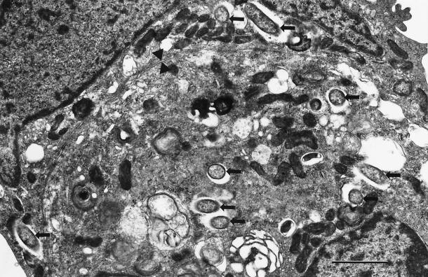
Owing to the potential role of the tick Amblyomma cooperi in the enzootic cycle of Rickettsia rickettsii, the etiologic agent of Brazilian spotted fever (BSF), this study evaluated infection by Rickettsia species in A. cooperi ticks collected from an area in Brazil where BSF is endemic. Among a total of 40 A. cooperi adult ticks collected in an area of BSF endemicity in the state of São Paulo, PCR analysis detected DNA of Rickettsia bellii in 16 ticks (40%), and 3 other ticks (7.5%) were positive for a previously unidentified spotted-fever-group (SFG) rickettsia. Cultivation in Vero cell cultures by the shell vial technique with individual A. cooperi ticks resulted in two isolates of R. bellii and one isolate genotypically characterized as an SFG rickettsia. The two R. bellii isolates were established in Vero cell cultures in the laboratory and were confirmed to be R. bellii by molecular analysis of the gltA and 17-kDa protein-encoding genes and by electron microscopic analysis. The SFG rickettsial isolate could not be stably passaged in cell culture in the laboratory, but molecular analysis of early passages suggested that it was closely related to Rickettsia parkeri, Rickettsia africae, and Rickettsia sibirica. These results do not support the role of A. cooperi in the ecology of R. rickettsii in the area studied, but they add two more species of rickettsiae to the poorly developed list of species occurring in ticks in South America.
Figures


References
-
- Bouyer, D. H., J. Stenos, P. C. Valdes, C. G. Moron, V. L. Popov, J. E. Zavala-Velazquez, L. D. Foil, D. R. Stothard, A. F. Azad, and D. H. Walker. 2001. Rickettsia felis: molecular characterization of a new member of the spotted fever group. Int. J. Syst. Evol. Microbiol. 51:339-347. - PubMed
-
- Burgdorfer, W. 1970. The hemolymph test. Am. J. Trop. Med. Hyg. 19:1010-1014. - PubMed
-
- Burgdorfer, W. 1988. Ecological and epidemiological considerations of Rocky Mountain spotted fever and scrub typhus, p. 33-50. In D. H. Walker (ed.), Biology of rickettsial diseases, vol. 1. CRC, Inc., Boca Raton, Fla.
-
- Cory, J., C. E. Yunker, J. A. Howarth, Y. Hokama, L. E. Hughes, L. A. Thomas, and C. M. Clifford. 1975. Isolation of spotted fever group and Wolbachia-like agents from field-collected materials by means of plaque formation in mammalian and mosquito cells. Acta Virol. 19:443-445. - PubMed
-
- Davis, G. E., and R. R. Parker. 1933. Additional studies on the relationship of the viruses of Rocky Mountain spotted fever and Sao Paulo exanthematic typhus. Pub. Health Rep. 48:1006-1011.
Publication types
MeSH terms
Grants and funding
LinkOut - more resources
Full Text Sources
