

doi: 10.1128/JCM.42.1.453-457.2004.
Development of a single-tube, cell lysis-based, genus-specific PCR method for rapid identification of mycobacteria: optimization of cell lysis, PCR primers and conditions, and restriction pattern analysis
Affiliations
- PMID: 14715804
- PMCID: PMC321669
- DOI: 10.1128/JCM.42.1.453-457.2004
Item in Clipboard
Development of a single-tube, cell lysis-based, genus-specific PCR method for rapid identification of mycobacteria: optimization of cell lysis, PCR primers and conditions, and restriction pattern analysis
J Clin Microbiol.
2004 Jan.
Abstract
A single-tube PCR method was developed for efficient identification of nontuberculous mycobacteria (NTM) and their environmental isolates in about 3 h without conventional DNA isolation. The following three steps were optimized or developed: (i). a simple, 6-min direct cell lysis protocol as a PCR prestep for generation of DNA-template, (ii). an improved Mycobacterium-specific PCR amplification protocol with a broader species specificity using newly designed primers targeting a 228-bp region of the 65-kDa heat shock protein (hsp) gene and optimal PCR amplification conditions, and (iii). a genus-specific restriction analysis of the PCR product for conclusive identification of the unknown NTM isolates.
Figures
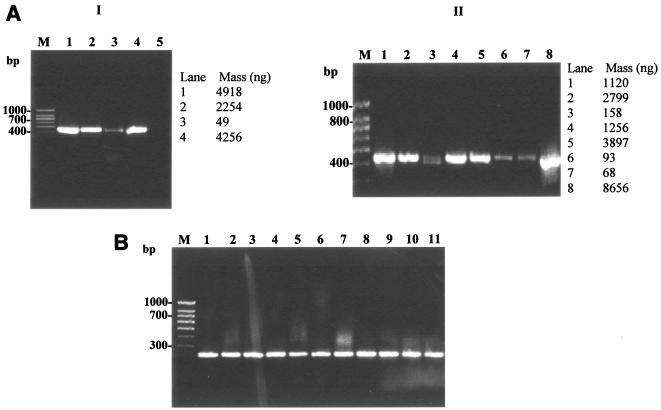
Optimization of cell lysis protocol for direct PCR-based detection of mycobacteria. (A) Optimization of chemical reagent for cell lysis as a prestep in hsp-based PCR amplification of M. smegmatis. Lanes 1 to 5, comparison of five different lysis reagents (reagents 1 through 5) containing various concentrations of SDS and Triton X-100 in Tris-EDTA buffer used in combination with thermal regime I (Table 1); lane 6, negative control (no template DNA). (B) Optimization of heating regimes for cell lysis of M. smegmatis using lysis reagent 5 (Table 1) in the hsp-based PCR. Lanes 1 to 5, comparison of thermal regimes 1 through 5 (Table 2); PCR amplification was based on mycobacterium-specific hsp gene with an expected 439-bp amplicon as described in the text. Lane M, 100-bp DNA size marker (PGC Scientifics).
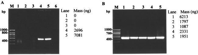
Evaluation of the optimized protocols for cell lysis and hsp-based PCR for mycobacteria. (A) Evaluation of the optimized cell lysis protocol (lysis reagent 5, thermal regime V) for hsp-based PCR amplification (439 bp) of mycobacterial reference strains and MWF isolates. Panel I: lanes 1 to 4, M. smegmatis, M. chelonae, M. immunogenum, and M. bovis; lane 5, no-template control. Panel II: lanes 1 to 8, M. smegmatis, M. chelonae, M. immunogenum, and Mycobacterium isolates M-JY1, M-JY2, M-JY3, M-JY4 and M-JY5; lane M, 100-bp DNA size marker (PGC Scientifics). PCR primers and conditions were the same as described in the legend of Fig. 1. (B) Evaluation of new hsp-based PCR primers and modified PCR conditions for genus-specific PCR amplification (228 bp) of mycobacteria. Lanes 1 to 3, M. smegmatis, M. chelonae, and M. immunogenum; lanes 4 to 11, Mycobacterium isolates M-JY1, M-JY2, M-JY3, M-JY4, M-JY5, M-JY6, M-JY7 and M-JY8; lane M, 100-bp DNA size marker (PGC Scientifics). Cells were lysed using the optimized direct cell lysis method as described for panel A, and the lysates were amplified using new PCR primers and conditions described in the text for amplification of a 228-bp PCR product of the hsp gene.
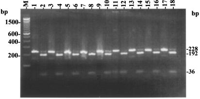
Confirmation of the mycobacterial origin of PCR amplicons (228 bp) based on NarI restriction patterns (192 and 36 bp). Shown are gel patterns for M. chelonae reference strain (lane 1 and 2) and for NTM field isolates M-JY1 (lanes 3 and 4), M-JY2 (lanes 5 and 6), M-JY3 (lanes 7 and 8), M-JY4 (lanes 9 and 10), M-JY5 (lanes 11 and 12), M-JY6 (lanes 13 and 14), M-JY7 (lanes 15 and 16), and M-JY8 (lanes 17 and 18); lane M, 100-bp DNA size marker (Invitrogen). For each isolate, the two lanes represent the original amplicon (8 μl) and its NarI restriction digestion product, respectively.
References
-
- Armand, M. O., M. Mestdagh, and F. Porteals. 2001. DNA isolation from chloroform/methanol-treated mycobacterial cells without lysozyme and proteinase K. BioTechniques 30:272-274. - PubMed
-
- Dailloux, M., C. Laurain, M. Weber, and P. Hartemann. 1999. Water and nontuberculosis mycobacteria. Water Res. 33:2219-2228.
-
- De Baere, T., R. de Mendonca, G. Claeys, G. Verschraegen, W. Mijs, R. Verhelst, S. Rottiers, L. Van Simaey, C. De Ganck, and M. Vaneechoutte. 2002. Evaluation of amplified rDNA restriction analysis (ARDRA) for the identification of cultured mycobacteria in a diagnostic laboratory. BMC Microbiol. 2:4-15. - PMC - PubMed
Publication types
MeSH terms
Associated data
- Actions
Grants and funding
LinkOut - more resources
Full Text Sources
Other Literature Sources
Molecular Biology Databases
