. 2004 Jan 7;24(1):8-23.
doi: 10.1523/JNEUROSCI.1650-03.2004.

Dopamine D1/D5 receptor modulates state-dependent switching of soma-dendritic Ca2+ potentials via differential protein kinase A and C activation in rat prefrontal cortical neurons

Affiliations

Dopamine D1/D5 receptor modulates state-dependent switching of soma-dendritic Ca2+ potentials via differential protein kinase A and C activation in rat prefrontal cortical neurons

Clint E Young et al. J Neurosci. .

Abstract

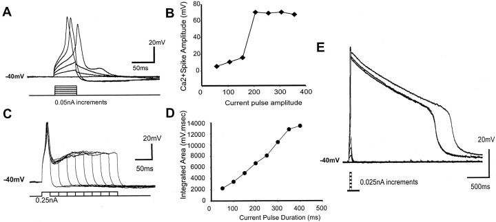
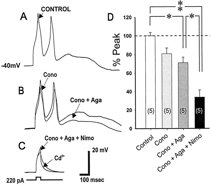
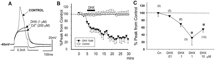
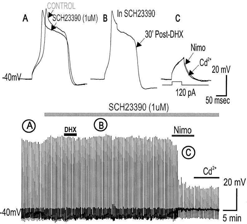
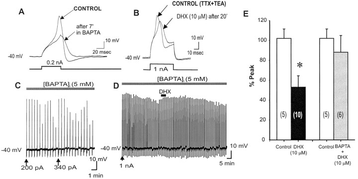
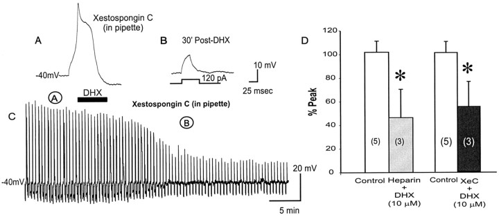
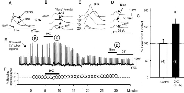
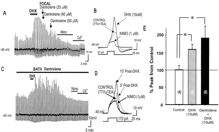
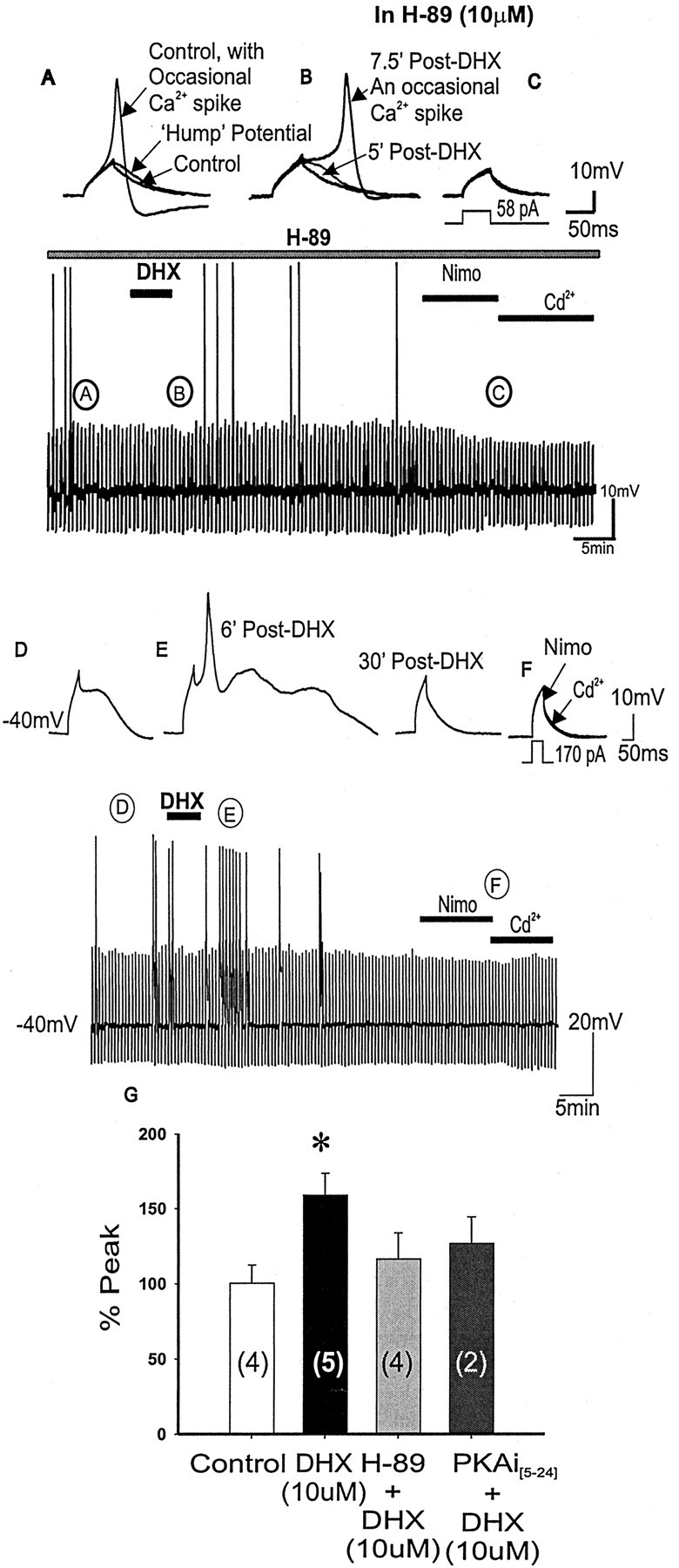
To determine the nature of dopamine modulation of dendritic Ca2+ signaling in layers V-VI prefrontal cortex (PFC) neurons, whole-cell Ca2+ potentials were evoked after blockade of Na+ and K+ channels. Soma-dendritic Ca2+ spikes evoked by suprathreshold depolarizing pulses, which could be terminated by superimposed brief intrasomatic hyperpolarizing pulses, are blocked by the L-type Ca2+ channel antagonist nimodipine (1 microM). The D1/D5 receptor agonist dihydrexidine (DHX) (0.01-10 microM; 5 min) or R-(+)SKF81291 (10 microM) induced a prolonged (>30 min) dose-dependent peak suppression of these Ca2+ spikes. This effect was dependent on [Ca2+]i- and protein kinase C (PKC)-dependent mechanisms because [Ca2+]i chelation by BAPTA or inhibition of PKC by bisindolymaleimide (BiM1), but not inhibition of [Ca2+]i release with heparin or Xestospongin C, prevented the D1-mediated suppression of Ca2+ spikes. Depolarizing pulses subthreshold to activating a Ca2+ spike evoked a nimodipine-sensitive Ca2+ "hump" potential. D1/D5 stimulation induced an N-[2-((o-bromocinamyl)amino)ethyl]-5-isoquinolinesulfonamide (H-89)- or internal PKA inhibitory peptide[5-24]-sensitive (PKA-dependent) transient (approximately 7 min) potentiation of the hump potential to full Ca2+ spike firing. Furthermore, application of DHX in the presence of the PKC inhibitor BiM1 or internal PKC inhibitory peptide[19-36] resulted in persistent firing of full Ca2+ spike bursts, suggesting that a D1/D5-PKA mechanism switches subthreshold Ca2+ hump potential to fire full Ca2+ spikes, which are eventually turned off by a D1/D5-Ca2+-dependent PKC mechanism. This depolarizing state-dependent, D1/D5-activated, bi-directional switching of soma-dendritic L-type Ca2+ channels via PKA-dependent potentiation and PKC-dependent suppression may provide spatiotemporal regulation of synaptic integration and plasticity in PFC.


Figures

Figure 1.
Figure 1.
Characterization of HVA Ca2+ spike in layers V-VI PFC pyramidal neurons using incremental intracellular depolarizing pulse amplitude and duration in the presence of TTX (1 μm) and TEA (200 μm). Three types of Ca2+ spikes were observed. A-D, Representative traces from neurons that fired single, double, or multiple Ca2+ spikes only, regardless of depolarizing pulse duration (33.7, 28.6, and 26.5% of the population, respectively). Note the sudden jump of spike amplitude (♦) when the all-or-nothing Ca2+ spike was elicited by 150 pA current pulse for this neuron (A, B), but a linear increase in the overall integrated area (•) of the Ca2+ spike and the ensuing post-spike plateau potential (C, D). E, In a small number of neurons (11.2% of the population), a small depolarizing pulse (50 msec) was sufficient to trigger a prolonged Ca2+ plateau that typically lasted for >1.5 sec (outlasting the short depolarizing pulse) followed by an abrupt repolarization. Incremental duration of suprathreshold pulse injected into these neurons did not elicit a longer Ca2+ plateau. These small number of neurons (n = 11) were present mostly in deep layer VI PFC pyramidal neurons.
Figure 2.
Figure 2.
Site of evoked Ca2+ spike electrogenesis determined by fast ISHPs. A, A short ISHP (-0.4nA, 10 msec) delivered in the midst of a suprathreshold depolarizing current pulse that previously evoked a robust Ca2+ spike, now effectively terminated the Ca2+ spike. This suggests that the Ca2+ spike was generated at a site relatively close to the somatic recording pipette. Such neurons were classified as having a “soma-proximal-basal dendritic site” of Ca2+ spike electrogenesis. B, In other neurons, longer-duration ISHPs (15-30 msec, -0.4 nA) delivered in the midst of a strong depolarizing current pulse were needed to terminate the Ca2+ spikes evoked. This suggests that the Ca2+ potential was generated relatively far from the somatic recording pipette. These neurons are classified as having a “distal site” of Ca2+ spike electrogenesis. C, D, In PFC neurons that elicit a prolonged Ca2+ plateau potential, in some (C), ISHPs (5-10 msec, -0.4 nA) abruptly terminate the Ca2+ plateau potentials (suggesting a “proximal site” of Ca2+ spike electrogenesis). On the other hand, there are also PFC neurons that can generate a prolonged Ca2+ plateau potential that failed to be terminated by strong and longer-duration ISHPs (D) (suggesting a distal site of Ca2+ spike electrogenesis).
Figure 3.
Figure 3.
Pharmacological dissection to show that L-type Ca2+ channels comprise the major component of the evoked Ca2+ spike. A, B, Representative traces showing a successive reduction of the suprathreshold evoked Ca2+ spike after the sequential application of a selective N-type Ca2+ channel blocker ω-conotoxin GVIA (Cono; 1 μm) (A) and a selective P-type Ca2+ channel blocker ω-agatoxin IVA (Aga; 100 nm) (B). D, E, Subsequent application of a specific L-type Ca2 blocker nimodipine (Nimo; 1 μm) greatly suppressed the Ca2 spike, whereas further exposure to cadmium (200 μm), a selective blocker of Ca2 channels, completely blocked any residual component of the Ca2+ potential so that only the membrane capacitative response remained. F, Histograms of group data showing that L-type Ca2+ channels account for a majority of the Ca2+ spike, and this is followed by N- and then P-type Ca2+ channels. *p < 0.05; **p < 0.001.
Figure 4.
Figure 4.
D1/D5R activation attenuates suprathreshold intrasomatic current evoked Ca2+ spike. A, Representative voltage traces showing a reduction of the suprathreshold evoked Ca2+ spike after D1/D5R activation by dihydrexidine (1 μm). Note that after a large suppression of the evoked Ca2+ spike by DHX, further addition of Cd2+ (200 μm) blocked a residual component of the Ca2+ potential so that only the membrane capacitative response remained. B, Open circles show no change in the amplitude of the Ca2+ spikes (evoked every 30 sec by a 50 msec intracellular depolarizing current pulse) over a typical recording period of >30 min, suggesting that under our recording condition, there was no run down of the evoked Ca2+ spike potentials. Filled triangles show that D1/D5R activation by DHX (10 μm) induced a prolonged (>30 min) reduction of the evoked Ca2+ spike. C, Graphic plots show that DHX dose dependently suppressed the peak but had little effect on the integrated area of the evoked Ca2+ spikes. Cn, Control.
Figure 5.
Figure 5.
Blocking D1/D5 receptors with SCH23390 prevents DHX-mediated suppression of high-threshold Ca2+ spikes. A, Application of the selective D1/D5 receptor blocker SCH23390 (1 μm) alone did not change the suprathreshold evoked Ca2+ spiking properties (peak and area) in PFC neurons. B, D1/D5 receptor activation by DHX (10 μm) in SCH23390 failed to attenuate the Ca2+ spike, suggesting that DHX-mediated attenuation of suprathreshold evoked Ca2 spikes requires D1/D5R activation. C, Subsequent application of a specific L-type Ca2 blocker nimodipine (10 μm) suppressed the Ca2 spike, whereas further exposure to cadmium (200 μm), a selective blocker of Ca2 channels, blocked a residual component of the Ca2+ potential so that only the membrane capacitative response remained. The bottom time-compressed traces indicate the time course of the response. D, Histograms of group data showing that D1/D5R activation (DHX alone) suppresses the Ca2+ spike but DHX application in the presence of SCH23390 prevents the suppression, suggesting that DHX-mediated suppression of suprathreshold evoked Ca2+ spikes depends on D1/D5R activation. Nimo, Nimodipine.
Figure 6.
Figure 6.
Intracellular chelation by BAPTA prevented D1/D5R suppression of high-threshold Ca2+ spikes. A, Ca2+ spikes evoked by suprathreshold depolarizing pulses a using BAPTA-filled pipette exhibit attenuated responses over time. C, Vertical lines are Ca2+ spikes (displayed in slower chart speed) that show that an increase in current pulse intensity is necessary to reestablish the same Ca2+ spike. This suggests that there may be a constitutively active intracellular Ca2+-dependent facilitation of evoked Ca2+ spikes. B, D, After achieving a steady-state evoked Ca2+ spike over time, D1/D5R activation by DHX (10 μm) failed to suppress the evoked Ca2+ spike recorded by the BAPTA-filled electrode. E, Histograms summarizing group data that illustrate that the suppression of the evoked Ca2+ spike was blocked with steady-state intracellular Ca2+ chelation by BAPTA. This suggests that D1/D5R suppression of suprathreshold-evoked Ca2+ spikes is Ca2+ dependent.
Figure 7.
Figure 7.
Blocking intracellular Ca2+ release from IP3R store by Heparin or Xestospongic C does not change the D1/D5R-mediated suppression of suprathreshold Ca2+ spikes. A, Presence of the selective IP3R inhibitor Xestospongin C in the recording pipette did not change the suprathreshold evoked Ca2+ spiking properties in PFC neurons. B, D1/D5R activation by DHX (10 μm) in the presence of Xestospongin C did not prevent D1/D5R-mediated suppression of the suprathreshold evoked Ca2+ spike, suggesting that D1/D5R-mediated suppression of suprathreshold evoked Ca2+ spikes does not depend on intracellular Ca2+ release from IP3R stores. C, Time course of the evoked Ca2+ spike response to DHX in the presence of Xestospongin C. A and B represent the time point at which representative traces were taken above. D, Histograms of group data showing that D1/D5R activation still suppresses the Ca2+ spike in the presence of a nonselective IP3R inhibitor (heparin) or a specific IP3R inhibitor (Xestospongin C), thus suggesting that D1/D5R-mediated suppression of suprathreshold evoked Ca2+ spikes does not depend on intracellular Ca2+ release from IP3R stores.
Figure 8.
Figure 8.
Inhibiting PKC activation by BiM1 or PKCi[19-36] prevented D1/D5R-mediated suppression of evoked suprathreshold Ca2+ spikes. A, Bath application of the selective PKC inhibitor BiM1 (25 nm) alone did not change the suprathreshold evoked Ca2+ spiking properties (peak and area) in PFC neurons. B, D1/D5R activation by DHX (10 μm) in the presence of BiM1 failed to attenuate the Ca2+ spike, suggesting that D1/D5R activation attenuates suprathreshold evoked Ca2 spikes through PKC activation. C, Subsequent application of a specific L-type Ca2 blocker nimodipine (10 μm) suppressed the Ca2 spike, whereas further exposure to cadmium (200 μm), a selective blocker of Ca2 channels, blocked a residual component of the Ca2+ potential so that only the membrane capacitative response remained. Bottom time-compressed traces indicate the time course of the response. D, Using PKCi [19-36] (10 μm) in the patch pipette did not change the suprathreshold evoked Ca2+ spiking properties in PFC neurons. E, D1/D5R activation by DHX (10 μm) with PKCi[19-36] (10 μm) in the patch pipette failed to attenuate the Ca2+ spike, suggesting that D1/D5R activation attenuates suprathreshold evoked Ca2 spikes through PKC activation. F, Subsequent application of a specific L-type Ca2 blocker nimodipine (10 μm) suppressed the Ca2 spike, whereas further exposure to Cd2+ (200 μm), a nonselective blocker of all Ca2 channels, blocked a residual component of the Ca2+ potential so that only the membrane capacitative response remained. Bottom time-compressed traces indicate the time course of the response. G, Histograms of group data showing that D1/D5R activation (DHX alone) suppresses the Ca2+ spike, but D1/D5R activation in the presence of a PKC inhibitor (BiM1) or PKCi[19-36] (10 μm) in the patch pipette prevents the suppression, suggesting that D1/D5R-mediated suppression of suprathreshold evoked Ca2+ spikes depends on PKC activation. *p < 0.05.
Figure 9.
Figure 9.
During suprathreshold depolarizations, D1/D5R activation transiently potentiates a secondary Ca2+ hump potential that led to spike firing. A, Representative voltage traces showing a single suprathreshold evoked Ca2+ spike (arrow). After D1/D5R activation, a Ca2+ hump potential emerges that trails the first Ca2+ spike. This Ca2+ hump potential eventually leads to spiking after 9′ post-D1/D5R activation (open arrow). Eventually, this potentiation subsides and completely disappears before the first Ca2+ spike is completely suppressed. B, Graphic representation of the peak amplitude of the first Ca2+ spike (filled circle) and the potentiated second Ca2+ spike (open circle) after brief D1/D5R activation by DHX for 5 min.
Figure 10.
Figure 10.
D1/D5R activation transiently potentiates a Ca2+ hump potential triggered by subthreshold depolarizing pulses. A, Ca2+ hump potential evoked by subthreshold depolarizing pulses was blocked by the L-type Ca2+ channel antagonist nimodipine (1 μm), suggesting that the Ca2+ hump potential is mediated by a low-threshold L-type Ca2+ channel subtype. B-D, Time course of the effects of D1/D5R stimulation to subthreshold Ca2+ hump potential. Note that occasional Ca2+ spikes are observed in the control. Representative traces from the control are shown (B). After DHX application, subthreshold hump potential now elicited a full Ca2+ spike (C), and the frequency of occurrence of full Ca2+ spikes is increased transiently (for ∼15 min). This enhancement was gradually reduced over time. After the potentiation had subsided, only the membrane capacitative response was evoked by the subthreshold depolarizing pulse. Further addition of nimodipine (1 μm) and cadmium (200 μm) (D) did not change the profile of the evoked response, suggesting that the spikes were recorded entirely from L-type Ca2+ channels. E, Time-compressed traces indicate the time course of the response. F, There was no change in the membrane resistance that was continuously being monitored by a hyperpolarizing (-50 to -150 pA, 100 msec) prepulse before each weak depolarizing pulse delivered to evoke a subthreshold Ca2+ hump potential. G, Histograms of group data showing that D1/D5R activation potentiated the subthreshold evoked Ca2+ hump potential to Ca2+ spike firing, resulting in an increase in Ca2+ spike amplitude after 5 min of DHX application. *p < 0.05.
Figure 11.
Figure 11.
Blocking intracellular Ca2+ release from ryanodine receptor stores by dantrolene does not change the transient D1/D5R-mediated potentiation of the subthreshold evoked Ca2+ spike. A-D, Time course of subthreshold evoked Ca2+ potential in response to DHX with internal Ca2+ release is blocked by either focal (A) or bath application (C) of dantrolene. D1/D5R activation led to a transient increase in peak amplitude and integrated area of the resulting evoked Ca2+ spike, suggesting that release of Ca2+ from internal stores is not required for the potentiation of the subthreshold spike (nor for the inevitable termination of the potentiation). Representative traces are shown for focal (B) and bath (D)-applied dantrolene. Note: After DHX application, the apparent increase in the downward deflection of vertical lines (A, C) below -40 mV represents an increase in the afterhyperpolarizing potential immediately after the Ca2+ spikes. E, Histogram showing that both DHX alone and DHX with dantrolene resulted in increased Ca2+ peak amplitude after 5 min post-DHX application, suggesting that Ca2+ release from intracellular stores is not required for this D1/D5R mediated potentiation. *p < 0.05. NIMO, Nimodipine.
Figure 12.
Figure 12.
Blocking PKA activation by H-89 or PKAi[5-24] prevented D1/D5R-mediated enhancement of the evoked subthreshold Ca2+ hump potential. A-C, Representative traces corresponding to the continuous time-compressed traces of evoked subthreshold Ca2+ potentials or occasional evoked Ca2+ spikes with application of the specific PKA inhibitor H-89 alone. Subthreshold Ca2+ potentials or occasional Ca2+ spikes are observed in the control state (A), suggesting that spike firing properties are unchanged with H-89 (10 μm) application. After DHX application (B), the frequency of Ca2+ spikes is unchanged and no potentiation is observed, suggesting that D1/D5R potentiation of subthreshold Ca2+ hump potentials depends on PKA activation. Further addition of nimodipine and cadmium (C) resulted in membrane capacitive responses. Bottom time-compressed traces indicate the time course of the response. D-F, When using PKAi[5-24] (10 μm) in the patch pipette, application of DHX resulted in blocking most of the expected potentiation by the D1 agonist (E); however, a few hump potentials could still be potentiated to Ca2+ spikes (E). Further addition of nimodipine and cadmium (F) resulted in membrane capacitive responses. G, Histograms summarizing group data showing that although D1/D5R activation (with DHX alone) potentiated subthreshold Ca2+ plateaus, D1/D5R activation in the presence of PKA inhibitor H-89 or PKAi[5-24] (10 μm) in the patch pipette did not potentiate the Ca2+ potentials, suggesting that D1/D5R-mediated potentiation depends on PKA activation. The bottom time-compressed traces indicate the time course of the response.
Figure 13.
Figure 13.
When blocking PKC activation by PKC inhibitory peptide[19-36], D1/D5R stimulation on subthreshold Ca2+ hump potential resulted in a long-lasting enhancement of the hump potential to evoked sustained Ca2+ spike firing. A-D, Representative traces corresponding to the continuous time-compressed traces of evoked subthreshold Ca2+ potentials or occasional evoked Ca2+ spikes with the specific PKC inhibitory peptide[19-36] in the patch pipette (D). After DHX application (A), the frequency of occurrence of evoked Ca2+ spikes is increased through an increase in the area of the hump potential, creating two Ca2+ spikes. Multiple Ca2+ spikes ride on the potentiated hump potential (B), resulting in four Ca2+ spikes. The potentiation is long lasting (>20 min), and the spike is blocked by addition of nimodipine (C), suggesting that the spikes were recorded from L-type Ca2+ channels. Further addition of cadmium blocked a small residual component of the Ca2+ potential so that only the membrane capacitative response remained, suggesting that the spikes were recorded mainly from L-type Ca2+ channels. E, Histograms summarizing group data showing that in the presence of PKCi[19-36], the D1/D5R-mediated potentiation of the subthreshold Ca2+ hump potentials begins ∼5 min post-DHX and led to a long-lasting enhancement and spike firing past 30 min post-DHX.
Figure 14.
Figure 14.
Schematic model that illustrates the state-dependent bi-directional D1/D5R modulation of L-type Ca2+ potentials through PKA potentiation and PKC suppression. Functional incoming synaptic signals to the distal dendrites and reaching the soma represent numerous temporally and spatially contiguous synaptic signals from diverse inputs. Such synaptic signals can be categorized as either strong or weak inputs, evoking suprathreshold Ca2+ spikes and plateaus or subthreshold hump potentials, respectively. A critical frequency of back-propagating Na+ spikes can evoke large, regenerative, voltage-gated Ca2+ spikes in the distal dendritic initiation zone (Schiller et al., 1997; Larkum et al., 1999a), whereas a single back-propagating Na+ spike generated in the axon facilitates the initiation of voltage-gated Ca2+ spikes when it is coincidental with synaptic input to the same distal dendritic site (Larkum et al., 1999b). For synaptic integration, the timing and location of evoked voltage-gated Ca2+ responses in dendrites by strong or weak inputs is important (Oakley et al., 2001). Nevertheless, an active participation of dendritic Ca2+ in amplifying distal inputs may occur mainly when powerful repolarizing dendritic K+ channels are suppressed (Gonzalez-Burgos and Barrionuevo, 2001). Dopamine D1/D5 receptor activation is known to suppress several subtypes of soma-dendritic K+ currents (Kitai and Surmeier, 1993; Nisenbaum et al., 1998; Dong and White, 2003), thus enabling the actions of dendritic Ca2+ potential to contribute actively in synaptic signal amplification and integration. A, Weak synaptic inputs lead to weak dendritic depolarizations that frequently evoke Ca2+ hump potentials that are subthreshold to evoking a suprathreshold Ca2+ spike (Seamans et al., 1997). B, D1/D5R activation results in a transient amplification or “boost” of the weak synaptic signal through a D1R-Gs-PKA pathway that potentiates L-type Ca2+ channels over time. C, The D1/D5R-PKA potentiation of the Ca2+ hump led to full Ca2+ spike firing via L-type Ca2+ channel activation in soma-proximal-basal dendrites. This Ca2+ spike firing is temporary, however, because it is eventually suppressed as greater Ca2+ influx activates a Ca2+-dependent D1-Gq-calcyon-PKC suppression of the L-type Ca2+ channel, thereby reducing Ca2+ influx (A). D, On the other hand, incoming strong synaptic inputs or back-propagating Na+ spikes can evoke full suprathreshold Ca2+ spikes. Optimal stimulation of D1/D5R via a [Ca2+]i-dependent D1-Gq-calcyon-PKC mechanism may lead to a differential suppression of the L-type Ca2+ channel-mediated Ca2+ spikes in various regions of the dendrites. This may serve functionally to “sharpen” incoming synaptic signals before they are integrated in the soma. Strong stimulation of D1/D5 receptor leads to severe suppression of dendritic Ca2+ spikes and may serve to prevent any synaptic signals from being integrated or amplified.

References

    1. Artalejo CR, Ariano MA, Perlman RL, Fox AP (1990) Activation of facilitation calcium channels in chromaffin cells by D1 dopamine receptors through a cAMP/protein kinase A-dependent mechanism. Nature 348: 239-242. - PubMed
    1. Artalejo CR, Rossie S, Perlman RL, Fox AP (1992) Voltage-dependent phosphorylation may recruit Ca2+ current facilitation in chromaffin cells. Nature 358: 63-66. - PubMed
    1. Baufreton J, Garret M, Rivera A, de la Calle A, Gonon F, Dufy B, Bioulac B, Taupignon A (2003) D5 (not D1) dopamine receptors potentiate burst-firing in neurons of the subthalamic nucleus by modulating an L-type calcium conductance. J Neurosci 23: 816-825. - PMC - PubMed
    1. Bekkers JM (2000) Distribution and activation of voltage-gated potassium channels in cell-attached and outside-our patches from large layer 5 cortical pyramidal neurons of the rat. J Physiol (Lond) 525: 611-620. - PMC - PubMed
    1. Berger B, Gaspar P, Verney C (1991) Dopaminergic innervation of the cerebral cortex: unexpected differences between rodents and primates. Trends Neurosci 14: 21-27. - PubMed

Publication types

MeSH terms

LinkOut - more resources