

Wnt signaling mutants have decreased dentate granule cell production and radial glial scaffolding abnormalities
- PMID: 14715945
- PMCID: PMC6729560
- DOI: 10.1523/JNEUROSCI.4071-03.2004
Wnt signaling mutants have decreased dentate granule cell production and radial glial scaffolding abnormalities
Abstract
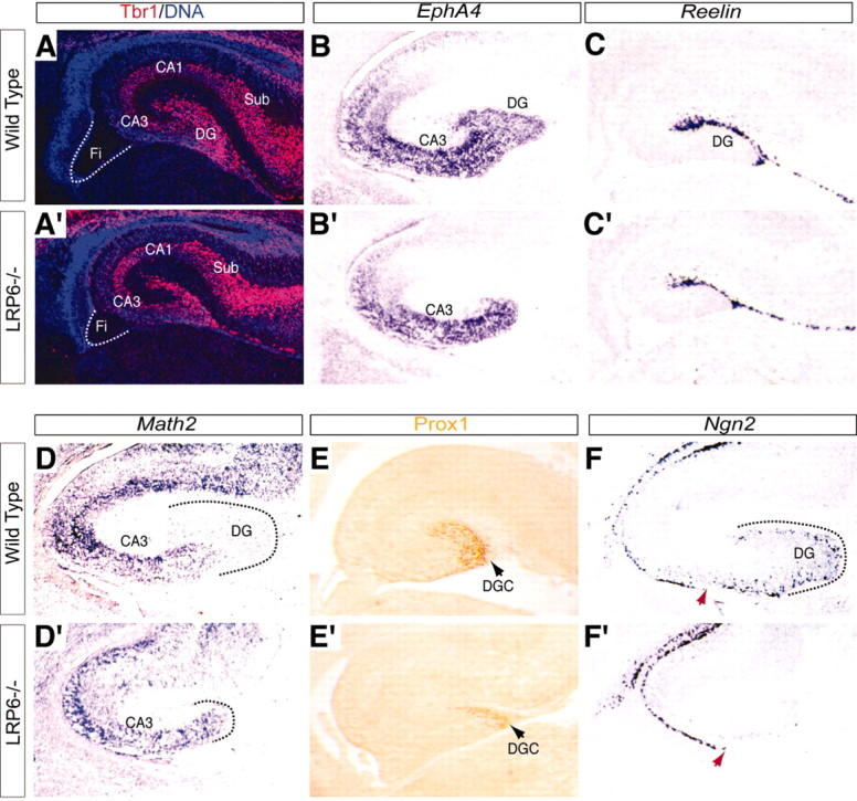
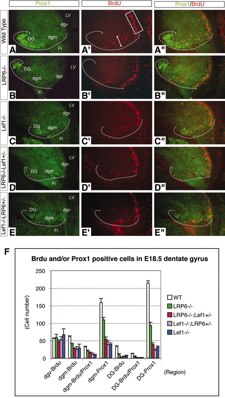
LRP6 mutant mice have generalized defects in the Wnt/beta-catenin signaling pathway because of the crucial function of LRP6 as a Wnt signaling co-receptor (Pinson et al., 2000). We examined the hippocampal phenotype of single LRP6 mutant mice as well as LRP6/Lef1 double mutant mice. LRP6 mutants had reduced production of dentate granule neurons and abnormalities of the radial glial scaffolding in the forming dentate gyrus. These defects were more severe with the addition of a single Lef1 null allele to an LRP6 null background. Pyramidal cell fields were unaffected in the LRP6, Lef1, or double mutants. The dentate defects were accompanied by decreased numbers of mitotic precursors in the migratory pathway to the dentate and in the displaced proliferative zone in the dentate itself. At earlier gestational ages, there was a reduction in the number of dentate granule cell progenitors in the dentate ventricular zone before the emigration of the earliest differentiated granule neurons and precursors to form the dentate anlage.
Figures


Similar articles
-
Hippocampus development and generation of dentate gyrus granule cells is regulated by LEF1.Development. 2000 Feb;127(3):469-82. doi: 10.1242/dev.127.3.469. Development. 2000. PMID: 10631168
-
Skeletal defects in ringelschwanz mutant mice reveal that Lrp6 is required for proper somitogenesis and osteogenesis.Development. 2004 Nov;131(21):5469-80. doi: 10.1242/dev.01405. Epub 2004 Oct 6. Development. 2004. PMID: 15469977
-
Morphogenesis of the dentate gyrus: what we are learning from mouse mutants.Dev Neurosci. 2005 Mar-Aug;27(2-4):93-9. doi: 10.1159/000085980. Dev Neurosci. 2005. PMID: 16046842 Review.
-
Unique expression patterns of cell fate molecules delineate sequential stages of dentate gyrus development.J Neurosci. 2000 Aug 15;20(16):6095-105. doi: 10.1523/JNEUROSCI.20-16-06095.2000. J Neurosci. 2000. PMID: 10934259 Free PMC article.
-
LDL receptor-related proteins 5 and 6 in Wnt/beta-catenin signaling: arrows point the way.Development. 2004 Apr;131(8):1663-77. doi: 10.1242/dev.01117. Development. 2004. PMID: 15084453 Review.
Cited by
-
Wnt signaling in development and disease.Neurobiol Dis. 2010 May;38(2):148-53. doi: 10.1016/j.nbd.2009.09.003. Epub 2009 Sep 16. Neurobiol Dis. 2010. PMID: 19765659 Free PMC article. Review.
-
GPCRs in stem cell function.Prog Mol Biol Transl Sci. 2013;115:175-216. doi: 10.1016/B978-0-12-394587-7.00005-1. Prog Mol Biol Transl Sci. 2013. PMID: 23415095 Free PMC article. Review.
-
Wnt7a regulates multiple steps of neurogenesis.Mol Cell Biol. 2013 Jul;33(13):2551-9. doi: 10.1128/MCB.00325-13. Epub 2013 Apr 29. Mol Cell Biol. 2013. PMID: 23629626 Free PMC article.
-
Dentate gyrus morphogenesis is regulated by β-catenin function in hem-derived fimbrial glia.Development. 2022 Nov 1;149(21):dev200953. doi: 10.1242/dev.200953. Epub 2022 Oct 21. Development. 2022. PMID: 36196585 Free PMC article.
-
The Wnt adaptor protein ATP6AP2 regulates multiple stages of adult hippocampal neurogenesis.J Neurosci. 2015 Mar 25;35(12):4983-98. doi: 10.1523/JNEUROSCI.4130-14.2015. J Neurosci. 2015. PMID: 25810528 Free PMC article.
References
-
- Altman J, Bayer SA (1990) Mosaic organization of the hippocampal neuroepithelium and the multiple germinal sources of dentate granule cells. J Comp Neurol 301: 325-342. - PubMed
-
- Amaral DG (1978) A Golgi study of cell types in the hilar region of the hippocampus in the rat. J Comp Neurol 182: 851-914. - PubMed
-
- Bagri A, Gurney T, He X, Zou YR, Littman DR, Tessier-Lavigne M, Pleasure SJ (2002) The chemokine SDF1 regulates migration of dentate granule cells. Development 129: 4249-4260. - PubMed
-
- Doetsch F, Caille I, Lim DA, Garcia-Verdugo JM, Alvarez-Buylla A (1999) Subventricular zone astrocytes are neural stem cells in the adult mammalian brain. Cell 97: 703-716. - PubMed
-
- Eckenhoff MF, Rakic P (1984) Radial organization of the hippocampal dentate gyrus: a Golgi, ultrastructural, and immunocytochemical analysis in the developing rhesus monkey. J Comp Neurol 223: 1-21. - PubMed
Publication types
MeSH terms
Substances
Grants and funding
LinkOut - more resources
Full Text Sources
Molecular Biology Databases