. 2004 Jan 7;24(1):127-37.
doi: 10.1523/JNEUROSCI.4591-03.2004.

Input-specific modulation of neurotransmitter release in the lateral horn of the spinal cord via adenosine receptors

Affiliations

Input-specific modulation of neurotransmitter release in the lateral horn of the spinal cord via adenosine receptors

Ruth E Brooke et al. J Neurosci. .

Abstract

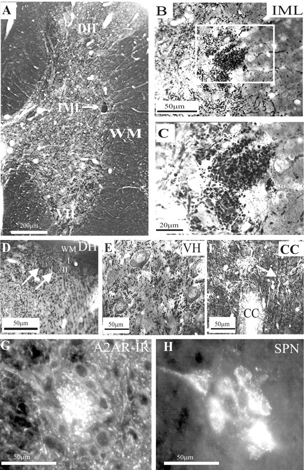
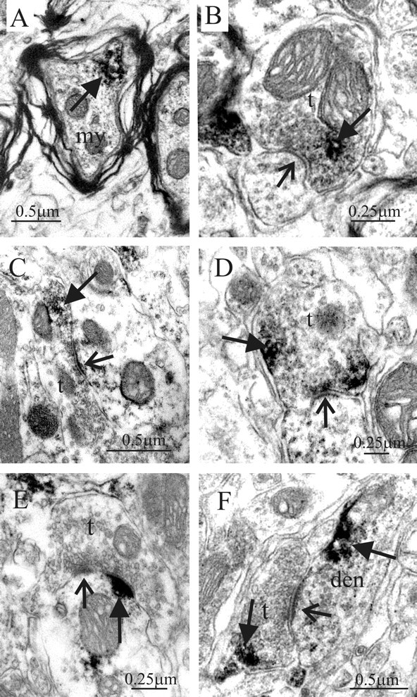
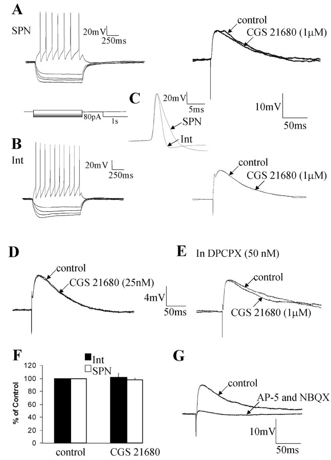
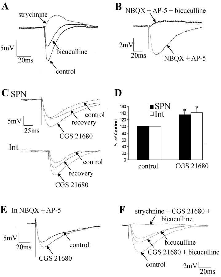
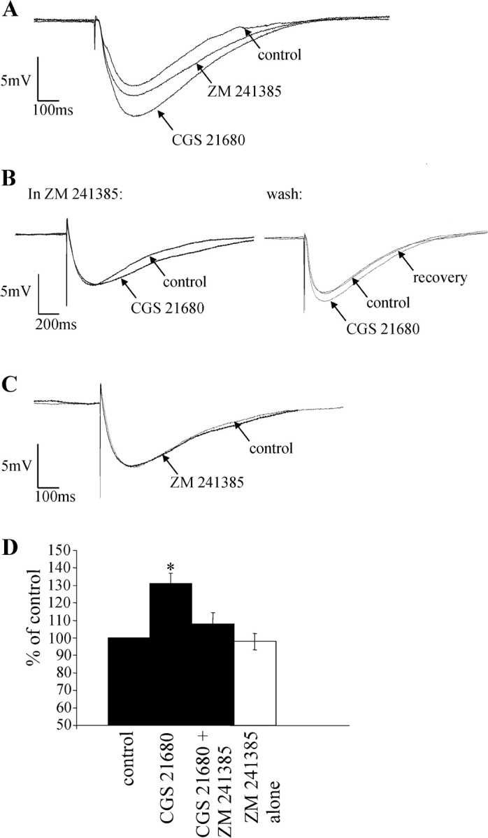
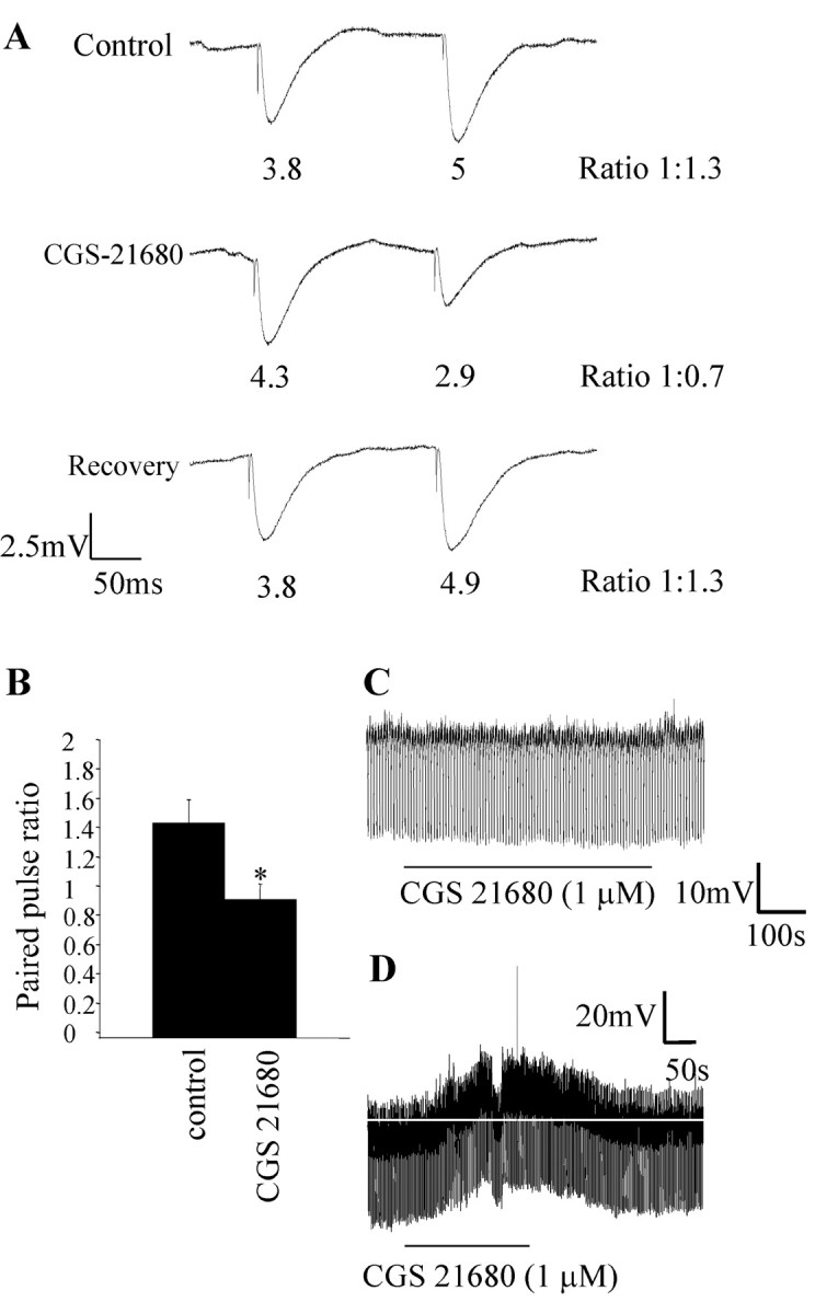
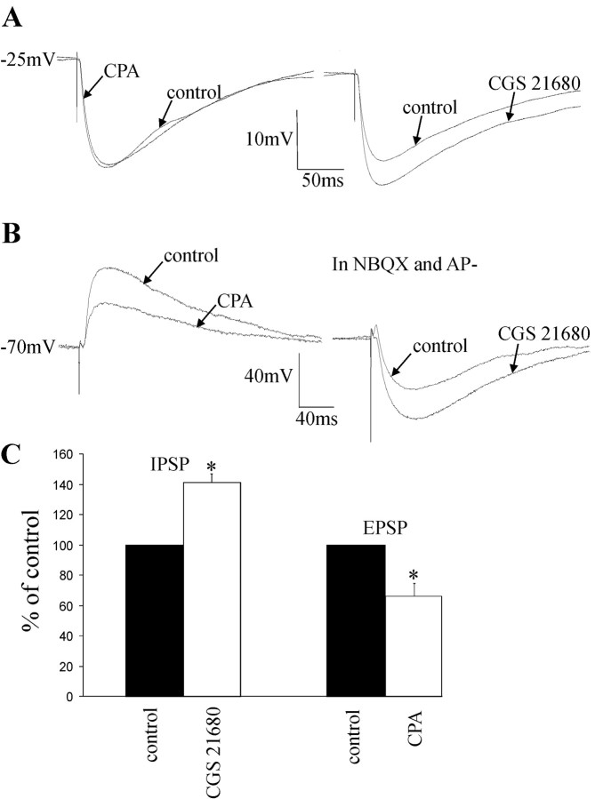
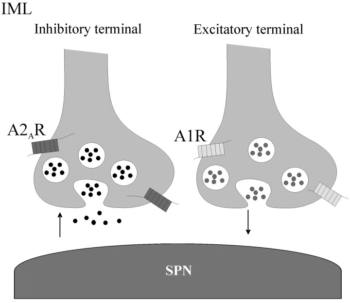
Activation of adenosine A2A receptors (A2ARs) in the CNS produces a variety of neuromodulatory actions dependent on the region and preparation examined. In autonomic regions of the spinal cord, A1R activation decreases excitatory synaptic transmission, but the effects of A2AR stimulation are unknown. We sought to determine the location and function of the A2ARs in the thoracic spinal cord, focusing on the intermediolateral cell column (IML). A2AR immunoreactivity was observed throughout the gray matter, with particularly dense immunostaining in regions containing sympathetic preganglionic neurons (SPNs), namely, the IML and intercalated nucleus. Electron microscopy revealed A2AR immunoreactivity within presynaptic terminals and in postsynaptic structures in the IML. To study the functional relevance of these A2ARs, visualized whole-cell patch-clamp recordings were made from electrophysiologically identified SPNs and interneurons within the IML. The A2AR agonist c2-[p-(carboxyethyl)phenethylamino]-5'-N-ethylcarboxyamidoadenosine (CGS 21680) had no significant effect on EPSPs but increased the amplitude of IPSPs elicited by stimulation of the lateral funiculus. These effects were attributable to activation of presynaptic A2ARs because CGS 21680 application altered the paired pulse ratio. Furthermore, neurons in the IML that have IPSPs increased via A2AR activation also receive excitatory inputs that are inhibited by A1R activation. These data show that activating A2ARs increase inhibitory but not excitatory transmission onto neurons in the IML. Simultaneous activation of A1Rs and A2ARs therefore could facilitate inhibition of the postsynaptic neuron, leading to an overall reduction of sympathetic nervous activity.


Figures

Figure 1.
Figure 1.
Specificity of A2AR primary antibody. A, Western blotting of rat thoracic spinal cord resulted in staining of a major band running at ∼45 kDa and a minor band of ∼62 kDa when detected with the anti-A2A receptor antibody. No staining of the membrane was obtained after preabsorption of the antiserum with the cognate peptide. B, Low magnification of A2AR immunoreactivity in the striatum. In accordance with previous reports, dense immunolabeling for the A2AR can be seen in the neuropil in the striatum. CPu, Caudate-putamen. C, A2AR immunoreactivity in the thoracic spinal cord visualized with DAB. Dense staining can be seen in the IML (arrow). D, No staining was observed after preincubation of the primary antibody with the immunogenic peptide. DH, Dorsal horn; VH, ventral horn.
Figure 2.
Figure 2.
Adenosine A2A receptor immunoreactivity in the thoracic spinal cord. A, Low magnification of A2AR immunoreactivity visualized with DAB. Labeling was observed throughout the spinal cord. Even at low magnification, a compact region of dense staining is clearly seen in the IML. Staining is also evident in the dorsal horn (DH) and ventral horn (VH). B, Low magnification of the IML. A compact region of punctate staining was observed in the IML adjacent to the white matter (WM), distinguishing this area from the surrounding neuropil. C, High magnification of the boxed area in B. At higher magnification, the punctate structures are clearly identifiable. D, In the dorsal horn, staining of fibers was dense in the superficial lamina, and labeled neuronal somata were also observed in lamina II (arrows). I, Lamina I; II, lamina II. E, In the ventral horn-labeled fibers, somata and proximal dendrites of large neurons were observed. F, In the vicinity of the central canal, labeling was observed in somata (arrow) as well as in presumptive fibers. G, High magnification of A2AR immunuoreactivity in the IML visualized with Cy3. A dense region of punctate staining can be observed in the IML. H, Sympathetic preganglionic neurons retrogradely labeled with Fluorogold in the same section as in G, viewed under UV illumination.
Figure 3.
Figure 3.
Electron microscopic localization of A2AR immunoreactivity in the IML. A, A2AR immunoreactivity (arrow) in a myelinated fiber (my) in the lateral funiculus. B, Presynaptic terminal (t) in the IML containing immunoreactivity for the A2AR (arrow, immunoreaction product). This terminal forms a synaptic contact (open arrow) with a dendritic structure. C, A large presynaptic terminal in the IML containing immunoreactivity (arrow) for the A2AR forms a synaptic contact (open arrow) with a dendritic profile. D, A2AR-immunoreactive terminal in synaptic contact (open arrow) with a dendritic structure. E, A2AR immunoreactivity (arrow) at the postsynaptic membrane of a synaptic junction (open arrow) where the presynaptic terminal is not labeled. F, Synaptic junction (open arrow) where both the presynaptic terminal and the postsynaptic dendritic (den) structure contained A2AR immunoreactivity (arrows).
Figure 4.
Figure 4.
The adenosine A2A receptor agonist CGS 21680 does not affect the amplitude of EPSPs elicited in SPNs and interneurons. A, Left, Voltage responses of an SPN to hyperpolarizing and depolarizing current pulses. A delayed return to resting potential at the end of the hyperpolarizing current pulse is evident, indicative of activation of an IA. Right, An EPSP was not significantly affected by CGS 21680 (1 μm). B, Left, Voltage responses of an interneuron (Int) to the same hyperpolarizing and depolarizing current pulses. At hyperpolarized potentials, a sag in the voltage response was seen, suggesting activation of an IH current. The action potential was short, and the AHP displayed distinct fast and slow phases. Right, Evoked EPSP elicited in the interneuron. The EPSP amplitudes are not significantly different from those elicited in the SPNs. In addition, CGS 21680 (1 μm) had no effect on the interneuronal EPSP. C, Comparison of the interneuronal and SPN action potentials and AHPs illustrating the shorter half-width of interneuronal action potentials. The SPN action potential shows the characteristic hump on the repolarization phase. D, EPSP elicited in an interneuron. CGS 21680 (25 nm) had no significant effect on the evoked EPSP. E, EPSP elicited in an SPN. CGS 21680 (1μm) administered in the presence of DPCPX had no significant effect on the evoked EPSP. F, Pooled data showing that CGS 21680 (1 μm) has no significant effect on EPSPs elicited in SPNs or interneurons. G, In another SPN, the EPSP was abolished by application of the excitatory amino acid antagonists NBQX (20 μm) and AP-5 (50 μm).
Figure 5.
Figure 5.
The adenosine A2A receptor agonist CGS 21680 increases the amplitude of IPSPs elicited in SPNs and interneurons. A, Evoked IPSP, which was partially blocked by bicuculline (10 μm) and abolished by the subsequent application of 1 μm strychnine. B, An IPSP in the presence of NBQX (20 μm) and AP-5 (50 μm) was completely blocked by the subsequent application of bicuculline (10 μm). C, Top, An IPSP elicited in the same SPN as shown in Figure 4A. CGS 21680 (1 μm) increased the amplitude of the IPSP from 5.5 to 10.8 mV. Bottom, IPSP elicited in the same interneuron (Int) as shown in Figure 4B. CGS 21680 (1 μm) increased the IPSP amplitude from 8.4 to 11.5 mV. D, Pooled data showing that 1 μm CGS 21680 increased the amplitude of IPSPs elicited in SPNs and interneurons and that this effect was not significantly different between the two types of neurons. *Significance at p > 0.05. E, IPSP elicited in an interneuron in the presence of the excitatory amino acid antagonists NBQX (20 μm) and AP-5 (50 μm). The IPSP remained in the presence of NBQX and AP-5, and this remaining component was increased in amplitude by CGS 21680 (1 μm). F, Evoked IPSP in an SPN, which was reduced in amplitude by bicuculline (10 μm). CGS 21680 (1 μm) enhanced the amplitude of the bicuculline-insensitive component, which was abolished by the subsequent application of strychnine (1 μm).
Figure 6.
Figure 6.
The effects of CGS 21680 are antagonized by the adenosine A2A receptor antagonist ZM 241385. A, IPSP elicited in an interneuron. CGS 21680 (1 μm) increased the amplitude of the IPSP, an effect that was antagonized by subsequent application of ZM 241385 (1 μm). B, Left, IPSP in an interneuron in the presence of 1 μm ZM 241385. After preincubation of ZM 241385, CGS 21680 (1 μm) failed to induce an increase in IPSP amplitude. Right, IPSP after washout of the drugs. The ability of CGS 21680 to increase the IPSP amplitude returned on washout of ZM 241385, indicating that preincubation of ZM 241385 blocked the effect of CGS 21680. C, Administration of ZM 241385 (1 μm) alone had no effect on the IPSP amplitude. D, Pooled data showing that CGS 21680 (1 μm) significantly increases IPSP amplitude and that this effect is antagonized by 1 μm ZM 241385. ZM 241385 alone has no effect on IPSP amplitude. *Significance at p > 0.05.
Figure 7.
Figure 7.
The effects of the adenosine A2A receptor agonist CGS 21680 are attributable primarily to a presynaptic site of action. A, Responses of an SPN to twin-pulse stimulation of the lateral funiculus in the control solution showed paired pulse facilitation; i.e., the response to the second stimulus was larger than the first. In 1 μm CGS 21680, the second response was smaller than the first response, with the ratio decreasing to 1:0.7. This change in ratio is indicative of a presynaptic site of action of CGS 21680. On washout of CGS 21680, the ratio returned to the control value of 1:1.3. B, Pooled data showing that 1 μm CGS 21680 altered the paired pulse ratio (n = 7). *Significance at p > 0.05. C, Hyperpolarizing current pulses (30 pA) were applied to an SPN (held at -65 mV), and 1 μm CGS 21680 was administered. There was no change in membrane potential or input resistance with CGS 21680 application. D, Example of a neuron in which a change in membrane potential was observed on administration of 1 μm CGS 21680. CGS 21680 caused a small depolarization (4 mV), which was not associated with a change in input resistance.
Figure 8.
Figure 8.
Selective modulation of excitatory and inhibitory synaptic inputs onto the same neuron by A1 and A2A adenosine receptors. A, Left, IPSP elicited in an SPN in control conditions and in the presence of CPA (100 nm). CPA had no effect on the amplitude of the IPSP. Right, After washout of CPA, CGS 21680 (1 μm) was applied, and an increase in the IPSP amplitude was obtained (20.6 to 26.6 mV). B, Left, EPSP elicited in an interneuron in control conditions and in the presence of CPA. CPA (100 nm) decreased the amplitude of the EPSP from 6.7 to 3.6 mV. Right, IPSP elicited in the same interneuron in the presence of NBQX (20 μm) and AP-5 (50 μm), observed at -20 mV. CGS 21680 (1 μm) increased the IPSP amplitude from 4.6 to 6.3 mV. C, Pooled data showing that 1 μm CGS 21680 increased the amplitude of evoked IPSPs, and 100 nm CPA decreased the amplitude of EPSPs elicited in the same neurons (n = 5). *Significance at p > 0.05.
Figure 9.
Figure 9.
The distribution of adenosine A1 and A2A receptors is input specific. This drawing summarizes the distribution of adenosine receptors in the IML of the thoracic spinal cord. A1Rs are located exclusively on excitatory terminals, whereas A2ARs are targeted to inhibitory synaptic inputs. Once activated, A2ARs increase neurotransmitter release, whereas A1Rs decrease release, from their respective terminals.

References

    1. Aden U, Halldner L, Lagercrantz H, Dalmau I, Ledent C, Fredholm BB (2003) Aggravated brain damage after hypoxic ischemia in immature adenosine A2A knockout mice. Stroke 34: 739-744. - PubMed
    1. Ambalavanar R, Morris R (1989) Fluoro-gold injected either subcutaneously or intravascularly results in extensive retrograde labelling of CNS neurones having axons terminating outside the blood-brain barrier. Brain Res 505: 171-175. - PubMed
    1. Anderson CR, Edwards SL (1994) Intraperitoneal injections of fluorogold reliably labels all sympathetic preganglionic neurons in the rat. J Neurosci Methods 53: 137-141. - PubMed
    1. Barraco RA, Martens KA, Parizon M, Normile HJ (1993) Adenosine A2a receptors in the nucleus accumbens mediate locomotor depression. Brain Res Bull 31: 397-404. - PubMed
    1. Blum D, Hourez R, Galas MC, Popoli P, Schiffmann SN (2003) Adenosine receptors and Huntington's disease: implications for pathogenesis and therapeutics. Lancet Neurol 2: 366-374. - PubMed

Publication types

MeSH terms

LinkOut - more resources