. 2004 Jan 12:4:2.
doi: 10.1186/1471-2180-4-2.

The single-stranded DNA-binding protein of Deinococcus radiodurans

Affiliations

The single-stranded DNA-binding protein of Deinococcus radiodurans

Julie Malia Eggington et al. BMC Microbiol. .

Abstract

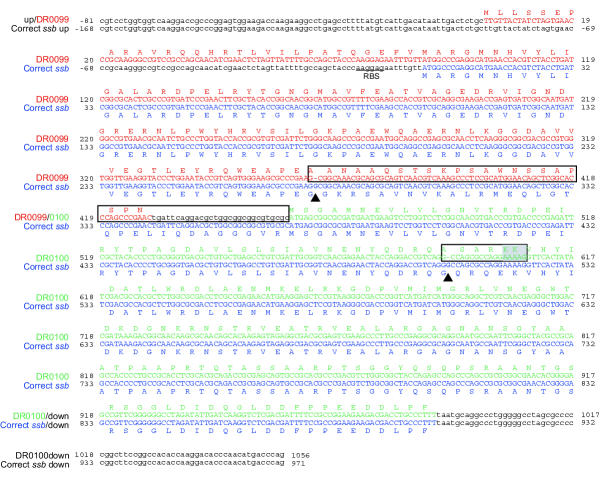
Background: Deinococcus radiodurans R1 is one of the most radiation-resistant organisms known and is able to repair an unusually large amount of DNA damage without induced mutation. Single-stranded DNA-binding (SSB) protein is an essential protein in all organisms and is involved in DNA replication, recombination and repair. The published genomic sequence from Deinococcus radiodurans includes a putative single-stranded DNA-binding protein gene (ssb; DR0100) requiring a translational frameshift for synthesis of a complete SSB protein. The apparently tripartite gene has inspired considerable speculation in the literature about potentially novel frameshifting or RNA editing mechanisms. Immediately upstream of the ssb gene is another gene (DR0099) given an ssb-like annotation, but left unexplored.

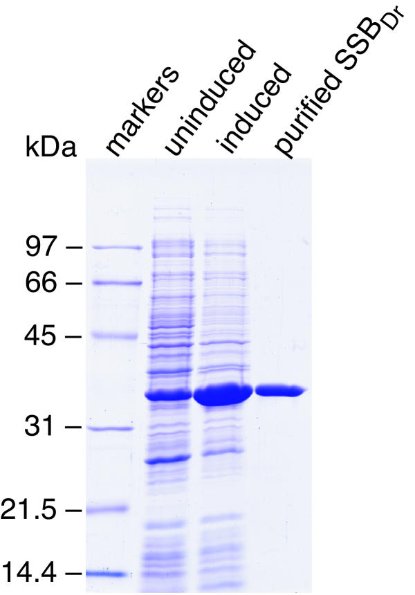
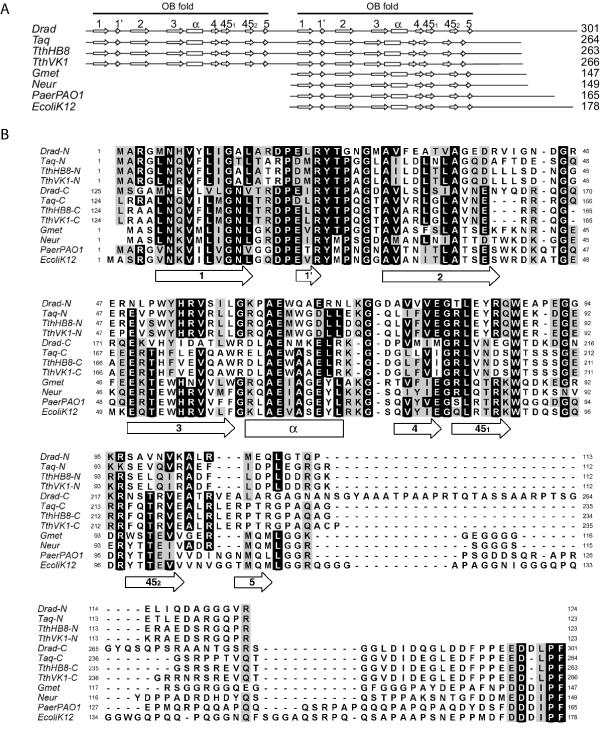
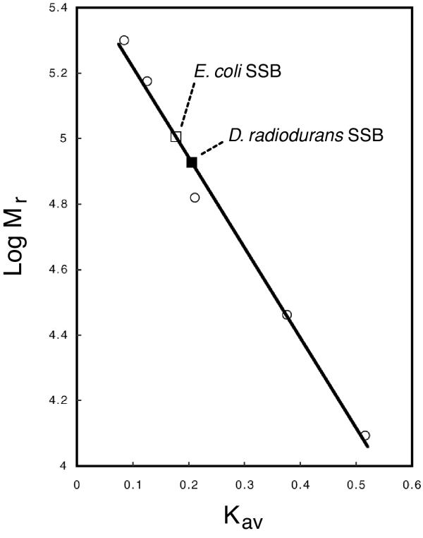
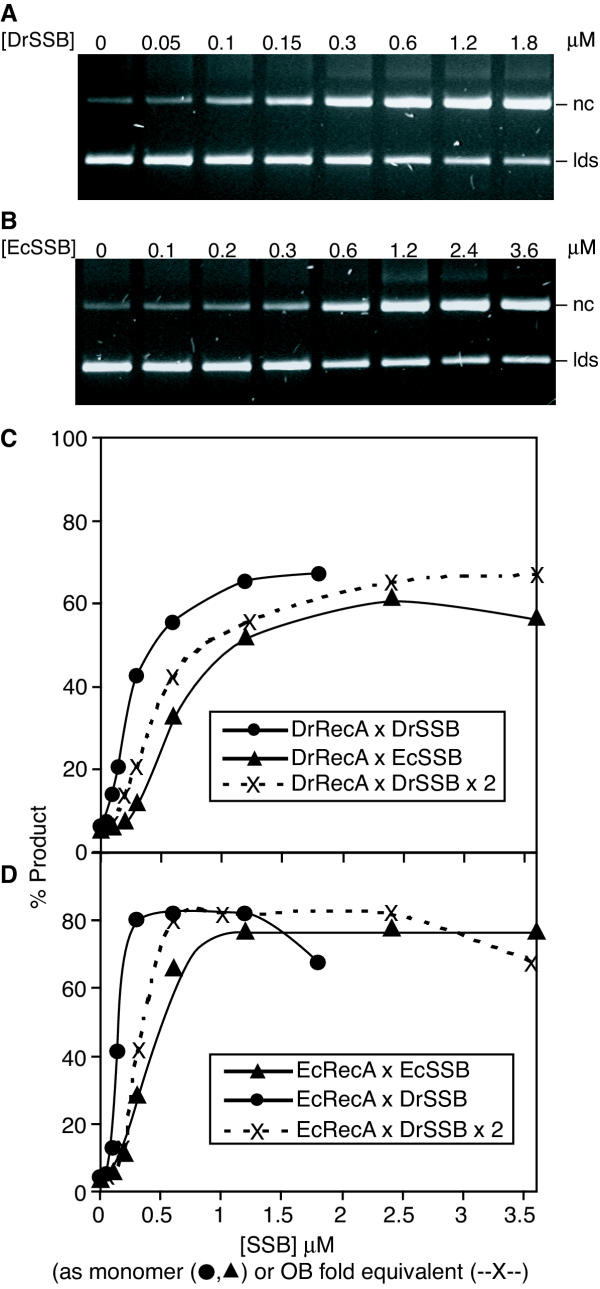
Results: A segment of the Deinococcus radiodurans strain R1 genome encompassing the ssb gene has been re-sequenced, and two errors involving omitted guanine nucleotides have been documented. The corrected sequence incorporates both of the open reading frames designated DR0099 and DR0100 into one contiguous ssb open reading frame (ORF). The corrected gene requires no translational frameshifts and contains two predicted oligonucleotide/oligosaccharide-binding (OB) folds. The protein has been purified and its sequence is closely related to the Thermus thermophilus and Thermus aquaticus SSB proteins. Like the Thermus SSB proteins, the SSBDr functions as a homodimer. The Deinococcus radiodurans SSB homodimer stimulates Deinococcus radiodurans RecA protein and Escherichia coli RecA protein-promoted DNA three-strand exchange reactions with at least the same efficiency as the Escherichia coli SSB homotetramer.

Conclusions: The correct Deinococcus radiodurans ssb gene is a contiguous open reading frame that codes for the largest bacterial SSB monomer identified to date. The Deinococcus radiodurans SSB protein includes two OB folds per monomer and functions as a homodimer. The Deinococcus radiodurans SSB protein efficiently stimulates Deinococcus radiodurans RecA and also Escherichia coli RecA protein-promoted DNA strand exchange reactions. The identification and purification of Deinococcus radiodurans SSB protein not only allows for greater understanding of the SSB protein family but provides an essential yet previously missing player in the current efforts to understand the extraordinary DNA repair capacity of Deinococcus radiodurans.


Figures

Figure 1
Figure 1
Nucleotide sequence of the D. radiodurans R1 ssb gene. The predicted amino acid sequence is shown above the nucleotide sequence for the DR0099 (red) and DR0100 (green)[4] and below for the corrected ssb (blue) sequences. Predicted non-coding regions are shown in lower case. The nucleotides omitted in the published sequence are shown in bold in the corrected ssb sequence and are indicated by triangles. The reading frames affected by the previous errors are shown in boxes. The translational frameshift region predicted by the earlier sequence is highlighted by a gray box. The putative Ribosomal Binding Site in the corrected ssb sequence is underlined. The sequences are numbered according to the first predicted initiation codon in the sequences (TTG in DR0099 and ATG in the corrected ssb). The DR0099 and DR0100 genes have accession number NC_001263. The corrected ssb sequence, reported here, has accession number AY293617.
Figure 2
Figure 2
Expression and purification of the D. radiodurans R1 SSB protein. Protein expression was obtained from the pEAW328 vector in BL21 Codon Plus (DE3) (Stratagene) E. coli cells. Proteins were examined on a standard SDS-polyacrylamide gel. Lanes are (1) molecular weight markers (BioRad, Low Range), as noted with the labels at left; (2) Whole cell extract of uninduced cells; (3) whole cell extract of cells after induction of the D. radiodurans ssb gene; (4) purified D. radiodurans R1 SSB protein.
Figure 3
Figure 3
Protein schematics and amino acid sequence alignments of D. radiodurans R1 SSB protein and other SSB proteins. A) Schematic representation of the D. radiodurans R1 SSB protein and other SSB proteins highlighting the OB fold regions. The Drad, Taq, TthHB8 and TthVK1 SSB proteins contain two OB folds each. The characteristic motifs that make up an OB fold are highlighted with open boxes/arrows and numbered. The Gmet, Neur, PaerPAO1 and EcoliK12 SSB proteins contain one OB fold each and align with both the N-terminal OB fold and the C-terminal OB fold of the proteins that contain two OB folds. Only the C-terminal alignments are shown. The structural assignments are according to the OB fold rule defined by Murzin [11,32]. B) Amino acid sequence alignment of D. radiodurans R1 SSB protein and other SSB proteins from closely and distantly related bacteria. The Drad, Taq, TthHB8 and TthVK1 SSB protein sequences are divided into N- and C-terminal fragments in order to highlight that each fragment contains an OB fold. The arrows indicate β-sheets and the rectangle indicates the α-helix contained in the secondary structure of the OB fold of SSB protein from E. coli strain K12 [11,32]. Sequence identity is shown by white fonts on black boxes, and sequence similarity is shown by black font on gray boxes. Sequence similarity is defined by the following amino acid groupings: STA, NEQK, NHQK, NDEQ, QHRK, MILV, MILF, HY, FYW. Abbreviations: Drad-N or -C: D. radiodurans strain R1, N- or C- terminal fragment; Taq-N or -C: T. aquaticus, N- or C- terminal fragment; TthHB8-N or -C: T. thermophilus strain HB8, N- or C- terminal fragment; TthVK1-N or -C: T. thermophilus strain VK1, N- or C- terminal fragment; Gmet: Geobacter metallireducens; Neur: Nitrosomonas europaea ATCC 19718; PaerPAO1: Pseudomonas aeruginosa PAO1. EcoliK12: E. coli strain K12.
Figure 4
Figure 4
Molecular weight approximation of the D. radiodurans SSB protein oligomer. Gel filtration of the standards and SSB proteins were performed as described in the Methods section using a S-200 Sephacryl HR column. The protein standards β-amylase (200 kDa), alcohol dehydrogenase (150 kDa), bovine serum albumin (66 kDa), carbonic anhydrase (29 kDa), and cytochrome c (12.4 kDa) were used to calibrate the column as shown by open circles. The elution volume of blue dextran was used to calculate the void volume and the total volume of the column was known. The best-fit line was generated of the log Mr of the protein standards versus Kav of the standards. D. radiodurans SSB protein and E. coli SSB protein samples were injected onto the column in independent experiments and their elution volumes were used to calculate their Kav. The position of D. radiodurans SSB protein on the standard curve is indicated by a closed square and the position of E. coli SSB protein on the standard curve is indicated by an open square.
Figure 5
Figure 5
DNA strand exchange reactions promoted by D. radiodurans RecA and E. coli RecA with SSB titrations. Reactions were carried out as described in the Methods section. Circular single-stranded DNA (css) was preincubated with either D. radiodurans or E. coli RecA. ATP and SSB protein (either D. radiodurans or E. coli SSB protein as indicated) were then added and incubated, followed by the addition of homologous linear double-stranded DNA (lds) which initiated the DNA three-strand exchange reaction. The nicked circular double-stranded DNA product (nc) is distinguishable by agarose gel electrophoresis and quantifiable. Panels A and B show the agarose gel electrophoresis results of reactions promoted by D. radiodurans RecA with various monomer concentrations of D. radiodurans SSB protein and E. coli SSB protein, respectively. These results are quantitated in panel C, with the data from reactions with D. radiodurans SSB (closed circles) and E. coli SSB (closed triangles) coming from Panels A and B respectively. Panel D shows the quantitated results of similar reactions promoted by E. coli RecA with D. radiodurans SSB (closed circle) and E. coli SSB (closed triangle) (agarose gel not shown). Since an SSBDr monomer has two OB folds and an SSBEc monomer has only one, the improved reactions seen in the reactions containing the former protein could simply reflect the higher effective concentration of OB folds when monomeric SSB concentrations are compared. In panels C and D, the dashed lines represent a plot in which the percentage reaction product generated in the reactions using SSBDr are plotted against the actual concentration of OB folds in these reactions (twice the actual concentration of D. radiodurans SSB monomers). The dashed lines (--X--) thus allow a direct comparison of the reactions observed with the D. radiodurans SSB protein with the reactions observed with the E. coli SSB protein (closed triangles). The production of nicked circular double-stranded DNA product is calculated as a percentage of total duplex DNA (the sum of linear double-stranded DNA substrate, nicked circular double-stranded DNA product and any network products near the well).

References

    1. Battista JR. Against all odds – the survival strategies of deinococcus radiodurans. Annu Rev Microbiol. 1997;51:203–224. doi: 10.1146/annurev.micro.51.1.203. - DOI - PubMed
    1. Makarova KS, Aravind L, Wolf YI, Tatusov RL, Minton KW, Koonin EV, Daly MJ. Genome of the extremely radiation-resistant bacterium Deinococcus radiodurans viewed from the perspective of comparative genomics. Microbiol Mol Biol Rev. 2001;65:44–79. doi: 10.1128/MMBR.65.1.44-79.2001. - DOI - PMC - PubMed
    1. Lohman TM, Ferrari ME. Escherichia coli single-stranded DNA-binding protein: multiple DNA-binding modes and cooperativities. Annu Rev Biochemistry. 1994;63:527–570. - PubMed
    1. White O, Eisen JA, Heidelberg JF, Hickey EK, Peterson JD, Dodson RJ, Haft DH, Gwinn ML, Nelson WC, Richardson DL, Moffat KS, Qin HY, Jiang LX, Pamphile W, Crosby M, Shen M, Vamathevan JJ, Lam P, McDonald L, Utterback T, Zalewski C, Makarova KS, Aravind L, Daly MJ, Minton KW, Fraser CM, et al. Genome sequence of the radioresistant bacterium Deinococcus radiodurans R1. Science. 1999;286:1571–1577. doi: 10.1126/science.286.5444.1571. - DOI - PMC - PubMed
    1. Lipton MS, Pasa-Tolic L, Anderson GA, Anderson DJ, Auberry DL, Battista JR, Daly MJ, Fredrickson J, Hixson KK, Kostandarithes H, Masselon C, Markillie LM, Moore RJ, Romine MF, Shen Y, Stritmatter E, Tolic N, Udseth HR, Venkateswaran A, Wong KK, Zhao R, Smith RD. Global analysis of the Deinococcus radiodurans proteome by using accurate mass tags. Proc Natl Acad Sci U S A. 2002;99:11049–11054. doi: 10.1073/pnas.172170199. - DOI - PMC - PubMed

Publication types

MeSH terms

LinkOut - more resources