Comparative Study
. 2004 Feb;141(3):477-87.
doi: 10.1038/sj.bjp.0705633. Epub 2004 Jan 12.

Octreotide regulates CC but not CXC LPS-induced chemokine secretion in rat Kupffer cells

Affiliations
Comparative Study

Octreotide regulates CC but not CXC LPS-induced chemokine secretion in rat Kupffer cells

Vassilis Valatas et al. Br J Pharmacol. 2004 Feb.

Abstract

Kupffer cells (KC) and lipopolysaccharide (LPS) interaction is the initial event leading to hepatic inflammation and fibrosis in many types of liver injury. We studied chemokine secretion by KC activated with LPS and the possible effect of the somatostatin analogue octreotide, in the regulation of this process. KC isolated from Sprague-Dawley rats were cultured in the presence of LPS added alone or with different concentrations of octreotide for 24 and 48 h, and chemokine production was assessed in culture supernatants by ELISA. CC chemokine mRNA expression was assessed by semiquantitative RT-PCR. Vehicle-stimulated KC produced a basal amount of CC and CXC chemokines. LPS-stimulated KC secreted significantly increased amounts of IL-8 (GRO/CINC-1) (P<0.001), MIP-2 (P<0.001), MCP-1 (P<0.001), and RANTES (P<0.01). Octreotide inhibited LPS-induced secretion of the CC chemokines MCP-1 (P<0.05) and RANTES (P<0.05), but not the CXC chemokines IL-8 (GRO/CINC-1) and MIP-2, in a concentration-dependent manner. Downregulation of basal and LPS-induced mRNA expression of the CC chemokines was also observed in the presence of octreotide. Pretreatment with phosphatidylinositol 3 (PI3)-kinase inhibitors reduced chemokine production by LPS-treated KC in both the mRNA and protein level. Furthermore, it prevented the octreotide inhibitory effect on LPS-induced chemokine secretion, indicating a possible involvement of the PI3-kinase pathway. In conclusion, these data demonstrate that chemokine secretion by KC can be differentially regulated by octreotide, and suggest that this somatostatin analogue may have immunoregulatory effects on resident liver macrophages. British Journal of Pharmacology (2004) 141, 477-487. doi:10.1038/sj.bjp.0705633


Figures
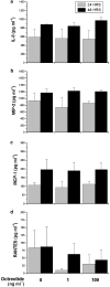
Figure 1
Figure 1
Octreotide has no effect on ‘basal' CXC and CC chemokine secretion by KC. KC cultures were treated with vehicle and different concentrations of octreotide, as described in ‘Experimental protocol for chemokine evaluation'. Chemokines were measured in culture supernatants by ELISA. (a) IL-8 (GRO/CINC-1), (b) MIP-2, (c) MCP-1, (d) RANTES. Values represent means±s.e.m. of three experiments.
Figure 2
Figure 2
Octreotide has no effect on LPS-induced CXC chemokine secretion by KC. KC cultures were treated with vehicle, LPS, and LPS/octreotide, as described in ‘Experimental protocol for chemokine evaluation'. Chemokines were measured in culture supernatants by ELISA. (a) IL-8 (GRO/CINC-1), (b) MIP-2. Values represent means±s.e.m. of 4–7 experiments. * represents significance from the LPS-treated group, *P<0.05, **P<0.01, ***P<0.001.
Figure 3
Figure 3
Octreotide inhibits LPS-induced MCP-1 secretion by KC. KC cultures were treated with vehicle, LPS, and LPS/octreotide, as described in ‘Experimental protocol for chemokine evaluation'. MCP-1 was measured in culture supernatants by ELISA. (a) 24 h incubation experiments, (b) 48 h incubation experiments, (c) percentage of octreotide-induced inhibition of MCP-1 production by LPS-stimulated KC cultures. Values represent means±s.e.m. of 4–7 experiments. * represents significance from the LPS-treated group, *P<0.05, **P<0.01, ***P<0.001.
Figure 4
Figure 4
Octreotide inhibits LPS-induced RANTES secretion by KC. KC cultures were treated with vehicle, LPS, and LPS/octreotide, as described in ‘Experimental protocol for chemokine evaluation'. RANTES was measured in culture supernatants by ELISA. (a) 24 h incubation experiments, (b) 48 h incubation experiments, (c) percentage of octreotide-induced inhibition of RANTES production by LPS-stimulated KC cultures. Values represent means±s.e.m. of 4–7 experiments. * represents significance from the LPS-treated group, *P<0.05, **P<0.01, ***P<0.001.
Figure 5
Figure 5
PI3-kinase inhibition suppresses LPS-induced MCP-1 secretion by KC. KC cultures were pretreated with wortmannin and stimulated with vehicle or LPS, as described in ‘Experimental protocol for chemokine evaluation'. Chemokines were measured in culture supernatants by ELISA. Values represent means±s.e.m. of three experiments. * represents significance from the LPS-treated group, *P<0.05.
Figure 6
Figure 6
PI3-kinase inhibition prevents octreotide suppression of LPS-induced MCP-1 secretion by KC. KC cultures were pretreated with wortmannin and stimulated with vehicle, LPS or LPS/octreotide, as described in ‘Experimental protocol for chemokine evaluation'. Chemokines were measured in culture supernatants by ELISA. Values represent means±s.e.m. of three experiments. * represents significance from the LPS-treated group, *P<0.05.
Figure 7
Figure 7
Octreotide inhibits MCP-1 mRNA expression by ‘resting' and LPS-stimulated KC. KC cultures were stimulated with vehicle, 1 μg ml−1 LPS (LPS), 0.1 ng ml−1 octreotide (OCT) or the combination of 1 μg ml−1 LPS with 0.1 ng ml−1 OCT for 24 h. Total mRNA was extracted from monolayer cultures following stimulation for 24 h, and MCP-1 mRNA expression was assessed with a semiquantitative RT–PCR, using specific primers for MCP-1 and 18s rRNA, as described under ‘RT–PCR'. The upper panel is the electrophoresis of the PCR products on agarose gels stained with ethidium bromide, and the lower panel is the densitometric analysis showing the relative expression of MCP-1 mRNA expressed as the MCP-1 to 18s ratios. Data are from a single experiment representative of at least three others.
Figure 8
Figure 8
Octreotide inhibits RANTES mRNA expression by ‘resting' and LPS-stimulated KC. KC cultures were stimulated with vehicle, 1 μg ml−1 LPS (LPS), 0.1 ng ml−1 octreotide (OCT) or the combination of 1 μg ml−1 LPS with 0.1 ng ml−1 octreotide for 24 h. Total mRNA was extracted from monolayer cultures following stimulation for 24 h, and RANTES mRNA expression was assessed with a semiquantitative RT–PCR, using specific primers for RANTES and 18s rRNA, as described under ‘RT–PCR'. The upper panel is the electrophoresis of the PCR products on agarose gels stained with ethidium bromide, and the lower panel is the densitometric analysis showing the relative expression of MCP-1 mRNA expressed as the MCP-1 to 18s ratios. Data are from a single experiment representative of at least three others.
Figure 9
Figure 9
PI3-kinase inhibition suppresses MCP-1 mRNA expression by KC. KC cultures were pretreated for 15 min with different concentrations of LY294002 (LY), and stimulated with 1 μg ml−1 LPS (LPS) for 24 h. Total mRNA was extracted from monolayer cultures following stimulation for 24 h and MCP-1 mRNA expression was assessed with a semiquantitative RT–PCR, using specific primers for MCP-1 and 18s rRNA, as described under ‘RT–PCR'. The upper panel is the electrophoresis of the PCR products on agarose gels stained with ethidium bromide, and the lower panel is the densitometric analysis showing the relative expression of MCP-1 mRNA expressed as the MCP-1 to 18s ratios. Data are from a single experiment representative of at least three others.
Figure 10
Figure 10
PI3-kinase inhibition suppresses RANTES mRNA expression by KC. KC cultures were pretreated for 15 min with different concentrations of LY294002 (LY), and stimulated with 1 μg ml−1 LPS (LPS) for 24 h. Total mRNA was extracted from monolayer cultures following stimulation for 24 h, and RANTES mRNA expression was assessed with a semiquantitative RT–PCR, using specific primers for MCP-1 and 18s rRNA, as described under ‘RT–PCR'. The upper panel is the electrophoresis of the PCR products on agarose gels stained with ethidium bromide, and the lower panel is the densitometric analysis showing the relative expression of RANTES mRNA expressed as the MCP-1 to 18s ratios. Data are from a single experiment representative of at least three others.

References

    1. AJUEBOR M.N., SWAIN M.G. Role of chemokines and chemokine receptors in the gastrointestinal tract. Immunology. 2002;105:137–143. - PMC - PubMed
    1. ANDOH A., HATA K., SHIMADA M., FUJINO S., TASAKI K., BAMBA S., ARAKI Y., FUJIYAMA Y., BAMBA T. Inhibitory effects of somatostatin on tumor necrosis factor-alpha-induced interleukin-6 secretion in human pancreatic periacinar myofibroblasts. Int. J. Mol. Med. 2002;10:89–93. - PubMed
    1. BAYON L.G., IZQUIERDO M.A., SIROVICH I., VAN ROOIJEN N., BEELEN R.H., MEIJER S. Role of Kupffer cells in arresting circulating tumor cells and controlling metastatic growth in the liver. Hepatology. 1996;23:1224–1231. - PubMed
    1. BONE-LARSON C.L., SIMPSON K.J., COLLETTI L.M., LUKACS N.W., CHEN S.C., LIRA S., KUNKEL S.L., HOGABOAM C.M. The role of chemokines in the immunopathology of the liver. Immunol. Rev. 2000;177:8–20. - PubMed
    1. BUKARA M., BAUTISTA A.P. Acute alcohol intoxication and gadolinium chloride attenuate endotoxin-induced release of CC chemokines in the rat. Alcohol. 2000;20:193–203. - PubMed

Publication types

LinkOut - more resources