Comparative Study
. 2004 Feb;123(2):109-19.
doi: 10.1085/jgp.200308935. Epub 2004 Jan 12.

Ionic currents mediated by a prokaryotic homologue of CLC Cl- channels

Affiliations
Comparative Study

Ionic currents mediated by a prokaryotic homologue of CLC Cl- channels

Alessio Accardi et al. J Gen Physiol. 2004 Feb.

Abstract

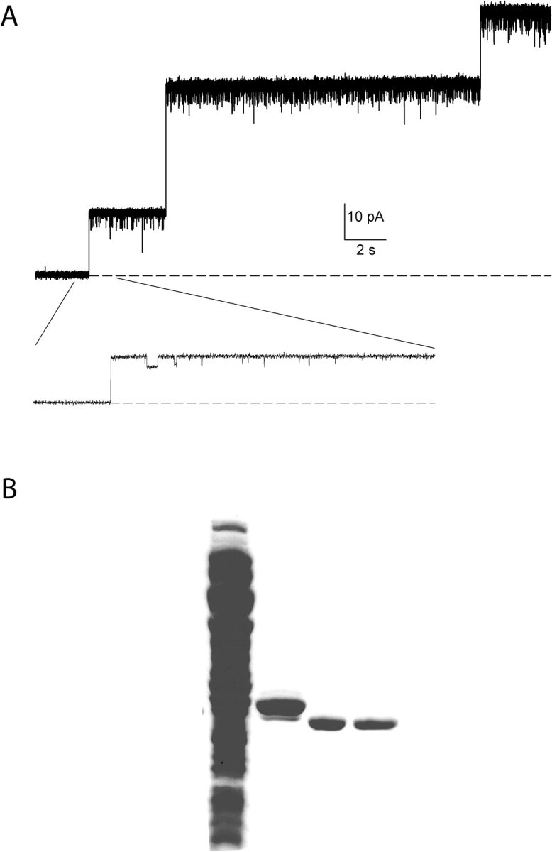
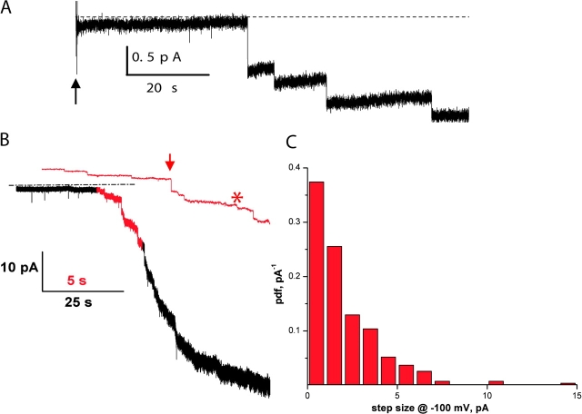
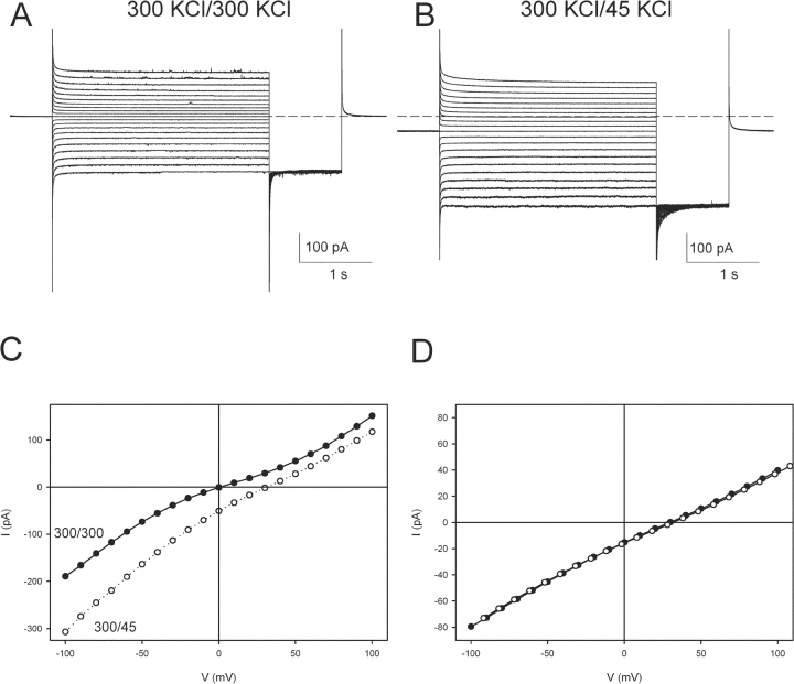
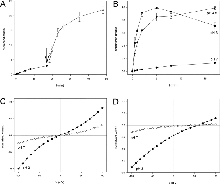
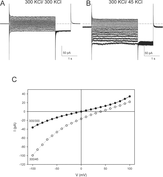
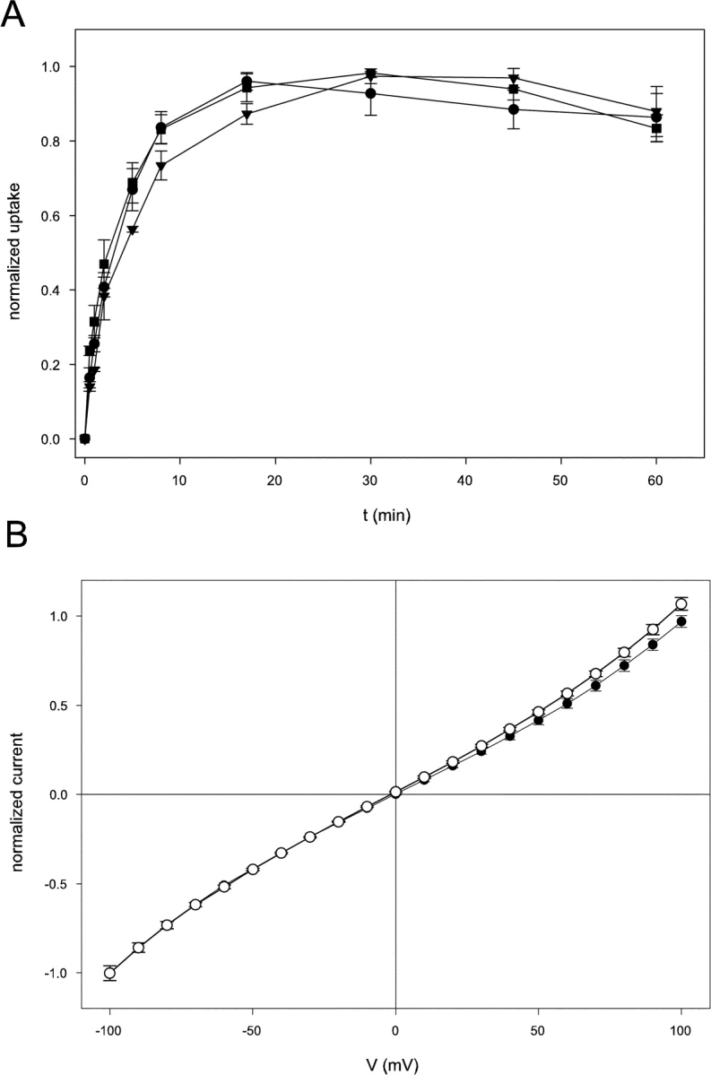
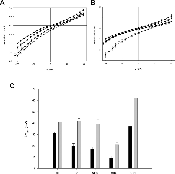
CLC-ec1 is an E. coli homologue of the CLC family of Cl- channels, which are widespread throughout eukaryotic organisms. The structure of this membrane protein is known, and its physiological role has been described, but our knowledge of its functional characteristics is severely limited by the absence of electrophysiological recordings. High-density reconstitution and incorporation of crystallization-quality CLC-ec1 in planar lipid bilayers failed to yield measurable CLC-ec1 currents due to porin contamination. A procedure developed to prepare the protein at a very high level of purity allowed us to measure macroscopic CLC-ec1 currents in lipid bilayers. The current is Cl- selective, and its pH dependence mimics that observed with a 36Cl- flux assay in reconstituted liposomes. The unitary conductance is estimated to be <0.2 pS. Surprisingly, the currents have a subnernstian reversal potential in a KCl gradient, indicating imperfect selectivity for anions over cations. Mutation of a conserved glutamate residue found in the selectivity filter eliminates the pH-dependence of both currents and 36Cl- flux and appears to trap CLC-ec1 in a constitutively active state. These effects correlate well with known characteristics of eukaryotic CLC channels. The E148A mutant displays nearly ideal Cl- selectivity.


Figures

F<sc>igure</sc> 1.
Figure 1.
Minuscule, debilitating porin contamination in CLC-ec1 prep. (A) Electrophysiological recording of crystal-quality CLC-ec1 prep incorporated into planar lipid bilayers. The recording is at 0 mV holding potential and cis/trans salt gradient conditions: 300 mM/40 mM KCl, pH 3. Liposomes contained CLC-ec1 at 30 μg/mg density. Dashed line represents zero-current level. Inset shows part of the trace on an expanded time scale. (B) Coomassie blue stained SDS gel illustrating the various steps in the CLC-ec1 purification procedure. Lane 1, crude membrane extract; lane 2, elution from Co column; lane 3, after digestion with LysC; lane 4, final preparation after anion exchange step.
F<sc>igure</sc> 2.
Figure 2.
CLC-ec1 fusions in lipid bilayers. (A) Anionic fusion-steps. Vesicles containing CLC-ec1 at protein to lipid ratio 50 μg/mg were added at the arrow to a bilayer under KCl-gradient conditions and zero-voltage holding potential. Solutions are cis HS side trans LS at symmetrical pH 3. Dashed line represents the zero current level. The trace was filtered at 50 Hz for display purposes. (B) Fusions at −100 mV. A recording similar to that in A was collected, except that protein density was 25 μg/mg and holding potential was −100 mV and filtering was at 500 Hz. Red portion of the trace is also shown on expanded scale, with 50 Hz filtering.
F<sc>igure</sc> 3.
Figure 3.
Macroscopic CLC-ec1 currents. (A) Macroscopic CLC-ec1 currents elicited with the following protocol: from a holding potential of 0 mV the voltage is stepped to −100 to +100 mV in 10-mV steps for 3 s. After a 1-s tail pulse to −100 mV the voltage is then returned to 0 mV. The solutions are symmetrical HS at pH 3.0 on both sides. Dashed line represents the zero current level. (B) Currents from the same bilayer as in A, when the solution on the trans side is LS at pH 3. Dashed line represents the zero current level. The stimulation protocol is the same as in A. (C) I-V curves of the traces shown in A (filled circles) and B (empty circles). (D) An IV curve was taken in a 300/45 mM KCl gradient as in B (filled circles). K+ was then replaced by NMG+ on both sides of the bilayer, and an IV curve was recorded (empty circles).
F<sc>igure</sc> 4.
Figure 4.
pH dependence of CLC-ec1. (A) Lowering the pH increases the 36Cl influx rate in CLC-ec1 containing liposomes. Uptake of 36Cl was monitored at pH 7.0 (filled circles), and the pH was then lowered to 4.5 (empty circles) by addition of H3PO4. The uptake is normalized to the total number of counts per sample. (B) Time course of 36Cl influx was followed at pH 7.0 (circles), 4.5 (triangles), and 3 (squares). Uptake is normalized to the steady-state uptake observed (equivalent to 20–30% of total counts, except at pH 3.0 where the uptake was roughly five- to tenfold lower). At pH 7.0 no steady-state was reached after as long as 4 h (not depicted). For normalization purposes the value at t = 17′ was assumed to be 13% of the steady-state value, in agreement with the results shown in A. (C) Normalized I-V curves of wildtype CLC-ec1 at pH 7.0 (empty circles) and pH 3.0 (filled circles) in symmetrical HS solutions. The I-Vs were normalized to the value of the currents at −100 mV in symmetrical HS solutions at pH 3.0 in the same bilayer. (D) Normalized I-V curves of wild-type CLC-ec1 at pH 7.0 (empty circles) and pH 3.0 (filled circles) with a salt gradient: HS solution on the cis side and LCL on the trans side. The I-V curves were normalized to the value of the currents at −100 mV in symmetrical HS solutions at pH 3.0 in the same bilayer.
F<sc>igure</sc> 5.
Figure 5.
Macroscopic currents of the E148A mutant. (A) Macroscopic E148A currents elicited with the same protocol as in 3A. The solutions are symmetrical HS at pH 3.0 on both sides. Dashed line represents the zero current level. (B) Currents from the same bilayer as in A, when the solution on the trans side is LS at pH 3. Dashed line represents the zero current level. The stimulation protocol is the same as in A. (C) I-V curves of the traces shown in A (filled circles) and B (empty circles).
F<sc>igure</sc> 6.
Figure 6.
pH dependence of the E148A mutant. (A) Time course of 36Cl influx for the E148A mutant was followed at pH 7.0 (circles), 4.5 (triangles), and 3 (squares). Uptake is normalized to the steady-state uptake observed (equivalent to 20–30% of total counts, except at pH 3.0 where the uptake was roughly five- to tenfold lower). (B) Normalized I-V curves of E148A CLC-ec1 at pH 7.0 (empty circles) and pH 3.0 (filled circles) in symmetrical HS solutions. The I-V curves were normalized to the value of the currents at −100 mV in symmetrical HS solutions at pH 3.0 in the same bilayer.
F<sc>igure</sc> 7.
Figure 7.
Selectivity of wild-type and E148A CLC-ec1. (A) Normalized I-Vs of wildtype CLC-ec1 in different ionic conditions in symmetrical pH 3. The solution on the cis side was HS; the trans side contained LS solution to which was added 255 mM KCl (circles), KBr (squares), K2SO4 (diamonds), or nothing (triangles). (B) Normalized I-Vs of E148A CLC-ec1 in the same conditions as in A (symbols are also the same). (C) Shifts in reversal potentials induced by test anions (Cl, Br, NO3 , SCN and SO4 =) in CLC-ec1 wildtype (black bars) and E148A (gray bars). The shifts, Vrev(X), were calculated for test anion X according to definition given in the text.

References

    1. Benz, R., and K. Bauer. 1988. Permeation of hydrophilic molecules through the outer membrane of gram-negative bacteria. Review on bacterial porins. Eur. J. Biochem. 176:1–19. - PubMed
    1. Chen, M.F., and T.Y. Chen. 2001. Different fast-gate regulation by external Cl− and H+ of the muscle-type ClC chloride channels. J. Gen. Physiol. 118:23–32. - PMC - PubMed
    1. Chen, T.-Y., and C. Miller. 1996. Nonequilibrium gating and voltage dependence of the ClC-0 Cl− channel. J. Gen. Physiol. 108:237–250. - PMC - PubMed
    1. Dutzler, R., E.B. Campbell, M. Cadene, B.T. Chait, and R. MacKinnon. 2002. X-ray structure of a ClC chloride channel at 3.0 Å reveals the molecular basis of anion selectivity. Nature. 415:287–294. - PubMed
    1. Dutzler, R., E.B. Campbell, and R. MacKinnon. 2003. Gating the selectivity filter in ClC chloride channels. Science. 300:108–112. - PubMed

Publication types