. 2004 Jan 19;164(2):255-66.
doi: 10.1083/jcb.200308132. Epub 2004 Jan 12.

Intraflagellar transport (IFT) cargo: IFT transports flagellar precursors to the tip and turnover products to the cell body

Affiliations

Intraflagellar transport (IFT) cargo: IFT transports flagellar precursors to the tip and turnover products to the cell body

Hongmin Qin et al. J Cell Biol. .

Erratum in

  • J Cell Biol. 2004 Mar 15;164(6):following 941

Abstract

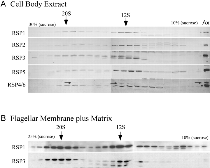
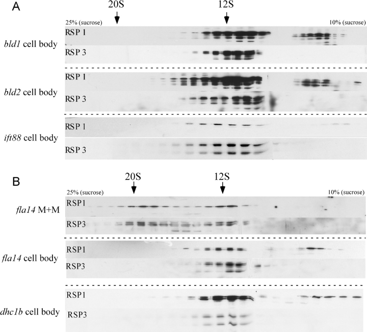
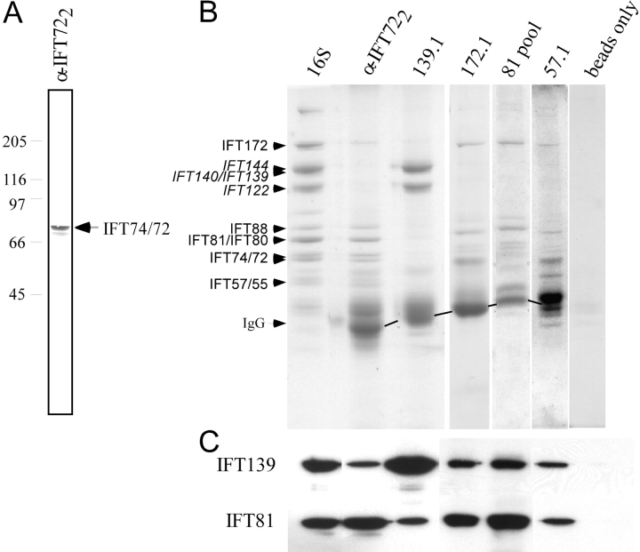
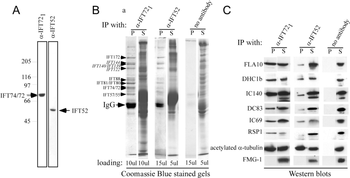
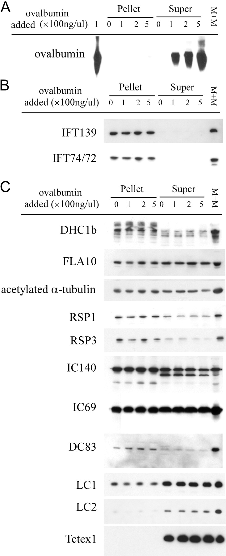
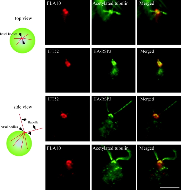
Intraflagellar transport (IFT) is the bidirectional movement of multisubunit protein particles along axonemal microtubules and is required for assembly and maintenance of eukaryotic flagella and cilia. One posited role of IFT is to transport flagellar precursors to the flagellar tip for assembly. Here, we examine radial spokes, axonemal subunits consisting of 22 polypeptides, as potential cargo for IFT. Radial spokes were found to be partially assembled in the cell body, before being transported to the flagellar tip by anterograde IFT. Fully assembled radial spokes, detached from axonemal microtubules during flagellar breakdown or turnover, are removed from flagella by retrograde IFT. Interactions between IFT particles, motors, radial spokes, and other axonemal proteins were verified by coimmunoprecipitation of these proteins from the soluble fraction of Chlamydomonas flagella. These studies indicate that one of the main roles of IFT in flagellar assembly and maintenance is to transport axonemal proteins in and out of the flagellum.


Figures

Figure 1.
Figure 1.
RSP1–6 from the cell body cosediment on gradients. (A) Fractions of a sucrose density gradient containing cell body extract were analyzed on immunoblots probed for RSPs as indicated. Note the 12S and 20S peaks of all RSPs examined. RSP3 migrates as a doublet due to phosphorylation; the 20S peak contains the upper phosphorylated band, characteristic of axonemal RSP3, whereas the 12S peak contains both forms. The RSP6 polyclonal antibody cross-reacts with RSP4 (arrows; these two RSPs are 48% identical [Curry et al., 1992]). RSP4 is only faintly visible in the 20S fraction. Ax, axoneme. (B) Gradient analysis of flagellar M+M was performed as in A. Both the 12S and 20S complexes are present in the M+M.
Figure 2.
Figure 2.
Negative-stained radial spokes from the 20S membrane plus matrix resemble mature spokes from the axoneme. Radial spokes from the 20S M+M were generally “T” shaped (panels 1–3), as are mature spokes (Yang et al., 2001); however, in some cases the stalk appeared to be coiled into a helix (panel 4). Occasionally the head formed three or four globular domains tethered to the stalk by thin filaments (panels 5–8). Bar, 20 nm.
Figure 3.
Figure 3.
Radial spoke proteins enter the flagellum as a 12S complex. (A) M+M fraction from regenerating wild-type cells were separated on 10–25% sucrose density gradients, fractions were separated on 8% gels and blots were probed for RSP1. During flagellar assembly the 12S complex predominates over the 20S complex. (B) Gradient analysis of the M+M of flagella from fla10 ts cells held at 32°C for 3 h (fla10 ts, 32°C), shows that the 20S form of radial spokes is more prevalent than the 12S form when IFT is blocked. The shift toward the 20S complex is observed to a lesser degree in fla10 ts cells at 22°C (fla10 ts, 22°C), at which temperature other minor effects of the mutation are apparent (Lux and Dutcher, 1991; Kozminski et al., 1995). Wild-type cell body extract is also shown (wt, 32°C) as a control. The blots were probed for RSP1.
Figure 4.
Figure 4.
Disassembled axonemal proteins accumulate in the membrane plus matrix when IFT is blocked. Flagella were isolated from fla10 ts cells incubated at 32°C for 0, 1, and 3 h or from wild-type cells treated with 1 mM IBMX for 75 min. Equal amounts of flagellar protein, or the M+M fraction from three times an equivalent amount of flagella, were processed for SDS-PAGE. (A) Coomassie blue–stained 10% gel of whole flagella and M+M. Molecular mass markers are indicated on the left. (B) Corresponding immunoblots probed with antibodies against IFT proteins and a variety of axonemal proteins (Table I). Note in B the disappearance of IFT proteins and the accumulation of axonemal proteins in the M+M of flagella of fla10 ts cells after 3 h at 32°C, but not in the M+M of flagella of IBMX-treated cells.
Figure 5.
Figure 5.
Retrograde IFT is required for the removal of the 20S radial spoke complex from the flagellum. (A) Cell body extracts of flagella-less cells were fractionated as in Fig. 3 and the blots were probed for RSP1 and -3. The 20S radial spoke complex present in wild-type cell bodies is absent from mutants without flagella - bld1, bld2, and ift88. (B) Gradient analysis of radial spoke complexes in M+M of fla14, which lacks retrograde IFT, shows a normal amount of the 20S complex (fla14 M+M). No 20S complex is present in the cell body of this mutant (fla14 cell body) or in dhc1b (dhc1b cell body), which also lacks retrograde IFT.
Figure 6.
Figure 6.
Intact IFT complex A and B can be immunoprecipitated separately. (A) Antibody α-IFT722 recognizes IFT74/72 on an immunoblot of the M+M. (B) Immunoprecipitates with antibodies against IFT polypeptides from M+M were analyzed on Coomassie blue–stained 8% gels. The antibodies used for immunoprecipitation are listed above the gels. The lane labeled 16S shows a sucrose density gradient fraction enriched for IFT polypeptides (Cole et al., 1998). IFT particle complex A (in italics) and B proteins are indicated. (C) Immunoblots of these gels show that IFT81and IFT139 are present in all the immunoprecipitates.
Figure 7.
Figure 7.
Complex A and B, IFT motors, and a variety of axonemal proteins coimmunoprecipitate with IFT particles. (A) Immunoblots of M+M probed with α-IFT721 and α-IFT52 show these antibodies recognize only the expected proteins. (B) The Coomassie blue–stained gel of immunoprecipitates obtained from M+M using α-IFT721 and α-IFT52 antibodies show complex A (in italics) and B proteins in the pellet (P), whereas most other proteins remain in the supernatant (S). A control was done without adding antibody (no antibody lanes); similar results were obtained using preimmune serum of α-IFT721. Pellets were resuspended in sample buffer equal to the supernatant volume. The loading volume is indicated at the bottom of each lane. (C) Corresponding immunoblots probed with antibodies against IFT motors FLA10 and DHC1b, and a variety of flagellar proteins as listed (Table I) show these proteins coprecipitated with IFT proteins. IFT52 is not shown because it is occluded by the IgG heavy chain.
Figure 8.
Figure 8.
Immunodepletion of IFT proteins from the membrane plus matrix fraction with α-IFT72 1 antibody precipitates a substantial fraction of IFT motors and several axonemal proteins. Ovalbumin was added to the M+M as a negative control at 100 to 500 ng/μl before immunoprecipitation with α-IFT721 antibody. Pellets were resuspended in a volume equal to the supernatants and equal volume loadings of pellets (Pellet), supernatants (Super), and M+M were resolved on a 10% gel. Immunoblots show that: (A) ovalbumin did not precipitate; (B) IFT polypeptides were immunodepleted from M+M (exemplified by IFT139 and IFT74/72; the rest of IFT particle proteins were similar; unpublished data); (C) immunoblots with antibodies against IFT motors FLA10 and DHC1b, and a variety flagellar proteins as listed (Table I) show these motor proteins and various flagellar proteins coprecipitated with IFT proteins. In contrast, LC2 and Tctex-1 did not precipitate.
Figure 9.
Figure 9.
IFT52 and RSP3 colocalize in the basal body area of the cell. (Left) Diagrams of the top and side views of a cell showing the position of basal bodies, flagella, and the four microtubule rootlets that emanate from the basal bodies. (Right) Immunofluorescence staining using antibodies as labeled. The first row shows the localization of FLA10 relative to the rootlet microtubules and basal bodies as shown in the diagram. Rows two and three show that the distribution of IFT52 and RSP3 surrounding the basal bodies generally overlap, though IFT52 covers a larger area. The fourth row illustrates the position of FLA10 relative to RSP3. Bar, 5 μm.

References

    1. Bell, S.E., A. Mavila, R. Salazar, K.J. Bayless, S. Kanagala, S.A. Maxwell, and G.E. Davis. 2001. Differential gene expression during capillary morphogenesis in 3D collagen matrices: regulated expression of genes involved in basement membrane matrix assembly, cell cycle progression, cellular differentiation and G-protein signaling. J. Cell Sci. 114:2755–2773. - PubMed
    1. Benashski, S.E., R.S. Patel-King, and S.M. King. 1999. Light chain 1 from the Chlamydomonas outer dynein arm is a leucine-rich repeat protein associated with the motor domain of the heavy chain. Biochemistry. 38:7253–7264. - PubMed
    1. Bloodgood, R.A., M.P. Woodward, and N.L. Salomonsky. 1986. Redistribution and shedding of flagellar membrane glycoproteins visualized using an anti-carbohydrate monoclonal antibody and concanavalin A. J. Cell Biol. 102:1797–1812. - PMC - PubMed
    1. Brazelton, W., C. Amundsen, C. Silflow, and P. Lefebvre. 2001. The bld1 mutation identifies the Chlamydomonas osm-6 homolog as a gene required for flagellar assembly. Curr. Biol. 11:1591–1594. - PubMed
    1. Cole, D.G. 2003. The intraflagellar transport machinery of Chlamydomonas reinhardtii. Traffic. 4:435–442. - PubMed

Publication types

Substances