. 2004 Mar;15(3):1479-86.
doi: 10.1091/mbc.e03-08-0622. Epub 2004 Jan 12.

Adenylyl cyclase G is activated by an intramolecular osmosensor

Affiliations

Adenylyl cyclase G is activated by an intramolecular osmosensor

Shweta Saran et al. Mol Biol Cell. 2004 Mar.

Abstract

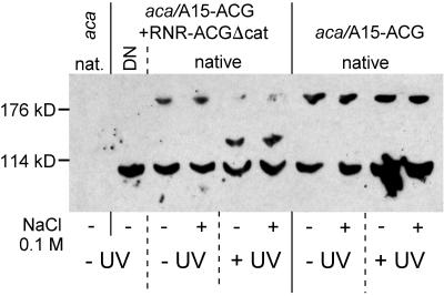
Adenylyl cyclase G (ACG) is activated by high osmolality and mediates inhibition of spore germination by this stress factor. The catalytic domains of all eukaryote cyclases are active as dimers and dimerization often mediates activation. To investigate the role of dimerization in ACG activation, we coexpressed ACG with an ACG construct that lacked the catalytic domain (ACGDeltacat) and was driven by a UV-inducible promoter. After UV induction of ACGDeltacat, cAMP production by ACG was strongly inhibited, but osmostimulation was not reduced. Size fractionation of native ACG showed that dimers were formed between ACG molecules and between ACG and ACGDeltacat. However, high osmolality did not alter the dimer/monomer ratio. This indicates that ACG activity requires dimerization via a region outside the catalytic domain but that dimer formation does not mediate activation by high osmolality. To establish whether ACG required auxiliary sensors for osmostimulation, we expressed ACG cDNA in a yeast adenylyl cyclase null mutant. In yeast, cAMP production by ACG was similarly activated by high osmolality as in Dictyostelium. This strongly suggests that the ACG osmosensor is intramolecular, which would define ACG as the first characterized primary osmosensor in eukaryotes.


Figures

Figure 1.
Figure 1.
Schematic of the ACG expression constructs. The constructs and protein structural domains are drawn according to scale except for the promoters (block arrows). The position and sequence of the peptide used to generate the αACG antibody is marked on the A15-ACG construct. The size of the proteins in amino acid residues is indicated.
Figure 2.
Figure 2.
Expression of ACG in S. cerevisiae. (A) Growth of the yeast cyr1 mutant TC41F2-1 (parent) and four clones (A1, A5, B1, and B5) transformed with the GAL1-ACG-ZZ fusion construct (cyr1/ACG) on standard yeast culture medium without cAMP. (B) Western blots of expressed ACG proteins. Lysates of Dictyostelium acg and aca/ACG cells, and of yeast cyr1 and cyr1/ACG cells were size fractionated by SDS-PAGE and immunoblotted with αACG antibodies. The size (in kilodaltons) of protein markers is indicated.
Figure 3.
Figure 3.
Osmoregulation of ACG in S. cerevisiae. (A) Time course of cAMP accumulation. cyr1/ACG cells were harvested from growth medium and resuspended in 5 mM MES, pH 6.0. Cells were stimulated with 1/10 volume of either 5 M NaCl or 1/5 volume of 5 M sorbitol. Reactions were quenched at the indicated time intervals by sixfold dilution in 60% methanol at -40°C. After cell rupture, cAMP levels were measured by isotope dilution assay. Means and SE of three experiments performed in triplicate are presented. (B) Dose-response curve. cyr1/ACG cells were stimulated with the indicated concentrations of NaCl. Reactions were quenched at 45 s after stimulation and assayed for cAMP. Means and SE of four experiments performed in triplicate are presented.
Figure 4.
Figure 4.
Effects of a truncated ACG protein on spore germination. Wild-type cells were transformed with a fusion construct of the psA prespore promoter and an ACG truncation that lacks the catalytic domain (psA-ACGΔcat). Wild-type and transformed spores either received a 45°C heat shock for 30 min or were left at 22°C before being incubated for 11 h in the presence and absence of 250 mM sucrose. Every 2 h, the ratio of spores to emerged amoebae was counted in a sample size of 100 cells. Means and SD of four experiments are presented.
Figure 5.
Figure 5.
Effect of conditional ACGΔcat expression on osmoregulation of ACG. aca cells were double transformed with constructs A15-ACG and RNR-ACGΔcat. The A15 and RNR promoters in these constructs direct constitutive and UV-inducible transcription, respectively. Effects of 100 mM NaCl on cAMP accumulation were measured in untransformed aca cells (A), uninduced transformants (B), and UV-induced transformants (C). Means and SE of three experiments performed in triplicate are presented.
Figure 6.
Figure 6.
Effects of high osmolality on ACG and ACGΔcat homo- and heterodimerization. aca cells, either single transformed with A15-ACG or double transformed with A15-ACG and RNRACGΔcat, were UV induced as indicated and subsequently treated with and without 100 mM NaCl for 5 min and lysed. The untransformed aca parent was lysed without pretreatment. Membranes were purified and membrane proteins were solubilized in 0.5% Triton X-100. For one sample of double-transformed cells, the membrane proteins were denatured (DN) by boiling in sample buffer containing 2% SDS. Equal amounts of native protein and denatured protein were loaded on nonreducing SDS-PAA gels and immunoblotted with αACG antibodies. The position of protein size markers is indicated.
Figure 7.
Figure 7.
Domain architecture of receptor adenylyl- and guanylyl cyclases. All sequences were retrieved from GenBank and the domain architecture was determined with the SMART program (Schultz et al., 1998). The size of the proteins in aa residues is indicated. The cyclase catalytic domains (AC or GC), kinase homology domain (KHD), ANP binding domain (ANP-R), coiled-coil, and transmembrane domains are drawn according to scale. Accession numbers are Homo sapiens ANPR: XM_113360; D. discoideum ACG: M87278; Trypanosoma brucei ESAG4: Q26721; T. brucei GRESAG4.3: X52121; T. brucei GRESAG4.4B: AF228602; Trypanosoma cruzi ADC1: AJ012096; T. cruzi zAC: AF040382; and Leishmania donovani RAC-A: U17042.

References

    1. Baldauf, S.L., Roger, A.J., Wenk-Siefert, I., and Doolittle, W.F. (2000). A kingdom-level phylogeny of eukaryotes based on combined protein data. Science 290, 972-977. - PubMed
    1. Bieger, B., and Essen, L.O. (2001). Structural analysis of adenylate cyclases from Trypanosoma brucei in their monomeric state. EMBO J. 20, 433-445. - PMC - PubMed
    1. Bilwes, A.M., Alex, L.A., Crane, B.R., and Simon, M.I. (1999). Structure of CheA, a signal-transducing histidine kinase. Cell 96, 131-141. - PubMed
    1. Blount, P., Sukharev, S.I., Moe, P.C., Schroeder, M.J., Guy, H.R., and Kung, C. (1996). Membrane topology and multimeric structure of a mechanosensitive channel protein of Escherichia coli. EMBO J. 15, 4798-4805. - PMC - PubMed
    1. Burke, D., Dawson, D., and Stearn, T. (2000). Methods in Yeast Genetics. Cold Spring Harbor, NY: Cold Spring Harbor Laboratory Press.

Publication types

LinkOut - more resources