. 2004 Apr;15(4):1580-90.
doi: 10.1091/mbc.e03-07-0544. Epub 2004 Jan 12.

TOGp, the human homolog of XMAP215/Dis1, is required for centrosome integrity, spindle pole organization, and bipolar spindle assembly

Affiliations

TOGp, the human homolog of XMAP215/Dis1, is required for centrosome integrity, spindle pole organization, and bipolar spindle assembly

Lynne Cassimeris et al. Mol Biol Cell. 2004 Apr.

Abstract

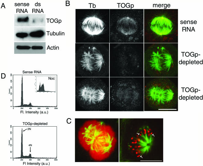
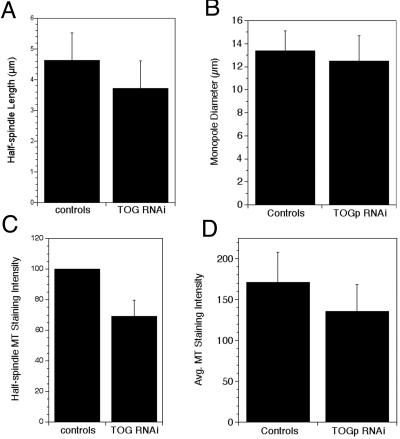
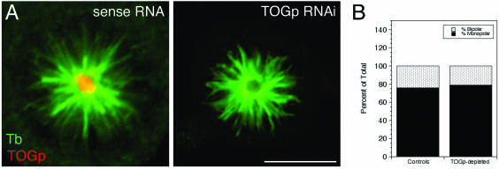
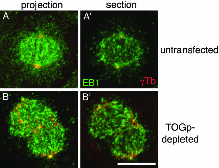
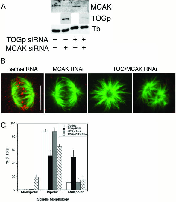
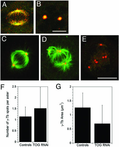
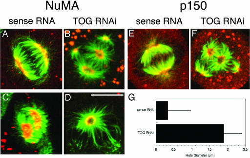
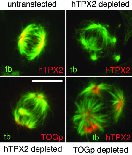
The XMAP215/Dis1 MAP family is thought to regulate microtubule plus-end assembly in part by antagonizing the catastrophe-promoting function of kin I kinesins, yet XMAP215/Dis1 proteins localize to centrosomes. We probed the mitotic function of TOGp (human homolog of XMAP215/Dis1) using siRNA. Cells lacking TOGp assembled multipolar spindles, confirming results of Gergely et al. (2003. Genes Dev. 17, 336-341). Eg5 motor activity was necessary to maintain the multipolar morphology. Depletion of TOGp decreased microtubule length and density in the spindle by approximately 20%. Depletion of MCAK, a kin I kinesin, increased MT lengths and density by approximately 20%, but did not disrupt spindle morphology. Mitotic cells lacking both TOGp and MCAK formed bipolar and monopolar spindles, indicating that TOGp and MCAK contribute to spindle bipolarity, without major effects on MT stability. TOGp localized to centrosomes in the absence of MTs and depletion of TOGp resulted in centrosome fragmentation. TOGp depletion also disrupted MT minus-end focus at the spindle poles, detected by localizations of NuMA and the p150 component of dynactin. The major functions of TOGp during mitosis are to focus MT minus ends at spindle poles, maintain centrosome integrity, and contribute to spindle bipolarity.


Figures

Figure 1.
Figure 1.
TOGp depletion results in multipolar spindle formation. (A) Immunoblot demonstrating decreased TOGp levels 48 h after transfection with dsRNA. Tubulin and actin levels were not changed by TOGp depletion. (B) Immunofluorescence micrographs of spindle morphology in TOGp-depleted cells 48 h after transfection. Images were collected as Z-stacks using confocal microscopy and converted to maximium intensity projections. Images were acquired and printed with identical settings. TOGp-depleted cells have multipolar spindles with prominent holes in the center of the spindle poles. (C) Chromosomes associate with all MT asters in TOGp-depleted cells. Left image, maximium intensity projection of TOGp-depleted cell double-labeled for tubulin (green) and DNA (red). Right image, single confocal optical section from TOGp-depleted spindle showing tubulin (green) and kinetochores (CREST staining, red). Arrows mark kinetochores associated with the ends of MT bundles. Bars (B and C), 10 μm. (D) FACS analysis of DNA content after TOGp depletion. Cells depleted of TOGp have normal DNA content, indicating that the multipolar spindles did not arise from a previous failed division. Inset, DNA content of cells incubated overnight with nocodazole. The peaks were used to assign the 2N and 4N peaks. In all cases, control cells were transfected with the sense RNA sequence alone.
Figure 3.
Figure 3.
TOGp does not make major contributions to MT length and density in the spindle. (A) Half spindle lengths measured from control (untreated or sense RNA transfected) and TOGp-depleted cells. Measured from 62 (control) and 90 (TOGp depleted) antitubulin-stained cells. Data from nine experiments, images collected by confocal microscopy. (B) Widths of monasters assembled in the presence of monastrol (100 μM). Data from three separate experiments, images collected by confocal microscopy (49 control and 58 TOGp-depleted cells). (C) MT staining intensity of half-spindles in control and TOGp-depleted cells. Data were normalized and pooled from three experiments. For each experiment the average intensity in controls (untreated and sense RNA transfected cells) was set to 100. Data from three experiments (102 control half spindles, 142 TOGp half spindles), images collected by wide-field microscopy. (D) MT staining intensity of monopolar spindles. Intensity was measured in a circle placed at the brightest region of the monoaster, adjacent to the central hole. Image stacks were collected by confocal microscopy, and a single optical section at the center of the monaster was used for quantitation. Data shown are from a representative experiment (15 control cells and 22 TOGp-depleted cells). All data are means ± SD.
Figure 2.
Figure 2.
Eg5 activity is required for multipolar spindle formation in TOGp-depleted cells. (A) Immunofluorescence micrographs (tubulin green, TOGp red) of monastrol-treated cells. Images are maximium intensity projections generated from confocal stacks. Scale bar, 10 μm. (B) Quantitation of the numbers of monopolar and bipolar spindles in monastrol-treated cells. No multipolar spindles were observed in TOGp-depleted cells and the percents of monopolar and bipolar spindles were identical to control cells (untreated or sense RNA-transfected cells).
Figure 4.
Figure 4.
Growing MT plus ends are abundant in TOGp-depleted cells. Growing MT plus ends were identified using an antibody to EB1 (green) and costained with antibodies against γ-tubulin to identify centrosomes (red). Images shown are maximium intensity projections and single optical sections from the center of the spindle. Growing MT plus ends were abundant in TOGp-depleted cells are were qualitatively of a density similar to that in untransfected spindles. Bar, 10 μm.
Figure 5.
Figure 5.
Simultaneous depletion of TOGp and MCAK results in bipolar and monopolar spindle formation. (A) Cell lysates were prepared 48 h after transfection with siRNA for TOGp, MCAK or both. Depletion of MCAK does not affect TOGp level and vice versa. Transfection of both TOG and MCAK siRNAs depletes cells of both proteins. Tubulin levels were unchanged by siRNA transfection. (B) Immunofluorescence micrographs of spindles assembled in MCAK- or TOGp/MCAK-depleted cells. MCAK localizes to kinetochores in cells transfected with sense RNA (red). MCAK depletion did not alter spindle morphology. Depletion of both TOGp and MCAK results in mono- and bipolar spindles. The TOG/MCAK-depleted cells were stained with rabbit antibodies to both TOGp and XKCM1 (MCAK). Bar, 10 μm. (C) Quantitation of spindle morphologies. Simultaneous depletion of TOGp and MCAK reverses the multipolar phenotype observed after TOGp depletion and results in a large increase in the number of monopolar spindles. Means ± SD from four experiments.
Figure 6.
Figure 6.
TOGp localizes to centrosomes in the absence of MTs and is required to maintain centrosome cohesion. (A and B) TOGp colocalizes with γ-tubulin in the presence (A) and absence (B) of MTs. (A and B) γ-tubulin (green) and TOGp (red). Cells in B were incubated in 30 μM nocodazole for 4 h before fixation. (C–F) Centrosomes fragment in the absence of TOGp. (C) Sense RNA–transfected cell; (D and E) cell fixed 48 h after transfection with siRNA to deplete TOGp. Cells are stained with antibodies to tubulin (C and D, green), TOGp (E, green), and γ-tubulin (C–E, red). In the TOGp-depleted cell, many asters contain multiple γ-tubulin foci. Bars, 10 μm. All images shown are maximium intensity projections. (E) The number of γ-tubulin foci per aster increased after TOGp depletion. The area of each γ-tubulin focus decreased after TOGp depletion. Data pooled from three experiments (34 asters in control cells [untransfected and sense RNA transfected] and 55 in TOGp-depleted cells).
Figure 7.
Figure 7.
TOGp depletion displaces MT minus ends from their tight focus at spindle poles. NuMA (A and C) and p150 (E) localized to a crescent-shaped structure at the spindle poles in sense RNA–transfected cells. In TOGp-depleted cells, NuMA (B and D) and p150 (F) localized to the edges of the holes in the center of each aster. (A, B, E, and F) are maximium intensity projections; (C and D) are single confocal sections showing end-on views of spindle poles. Panel C was generated from a 180° 3D projection tilted to view the spindle poles in a sense RNA–transfected cell. The NuMA crescent completely covers the front spindle pole. In other untransfected cells or cells transfected with sense RNA, small holes were sometimes observed in NuMA staining. Tubulin shown in green; NuMA or p150 shown in red. Bar, 10 μm. (G) The diameter of the central hole in the aster was ∼5 times wider after TOGp depletion.
Figure 8.
Figure 8.
hTPX2 and TOGp localize to the spindle independently of each other. Cells were transfected with siRNAs to deplete hTPX2 or TOGp, as marked. hTPX2 is undetectable in a large number of cells 36–40 h after siRNA transfection. Depletion of hTPX2 resulted in multipolar spindles, but TOGp remained localized to the centrosomes of these cells. hTPX2 retained spindle pole localization in the absence of TOGp. Bar, 10 μm.

References

    1. Becker, B.E., Romney, S.J., and Gard, D.L. (2003). XMAP215, XKCM1, NuMA and cytoplasmic dynein are required for the assembly and organization of the transient microtubule array during maturation of Xenopus oocytes. Dev. Biol. 261, 488-505. - PubMed
    1. Bellanger, J.M., and Gonczy, P. (2003). TAC-1 and ZYG-9 form a complex that promotes microtubule assembly in C. elegans embryos. Curr Biol. 13, 1488-1498. - PubMed
    1. Bornens, M. (2002). Centrosome composition and microtubule anchoring mechanisms. Curr. Opin. Cell Biol. 14, 25-34. - PubMed
    1. Cassimeris, L., Gard, D., Tran, P.T., and Erickson, H.P. (2001). XMAP215 is a long thin molecule which does not increase microtubule stiffness. J. Cell Sci. 114, 3025-3033. - PubMed
    1. Charrasse, S., Schroeder, M., Gauthier-Rouviere, C., Ango, F., Cassimeris, L., Gard, D.L., and Larroque, C. (1998). The TOGp protein is a new human microtubule-associated protein homologous to the Xenopus XMAP215. J. Cell Sci. 111, 1371-1383. - PubMed

Publication types

MeSH terms

LinkOut - more resources