. 2004 Mar;15(3):973-81.
doi: 10.1091/mbc.e03-07-0528. Epub 2004 Jan 12.

Fibronectin matrix assembly regulates alpha5beta1-mediated cell cohesion

Affiliations

Fibronectin matrix assembly regulates alpha5beta1-mediated cell cohesion

Elizabeth E Robinson et al. Mol Biol Cell. 2004 Mar.

Abstract

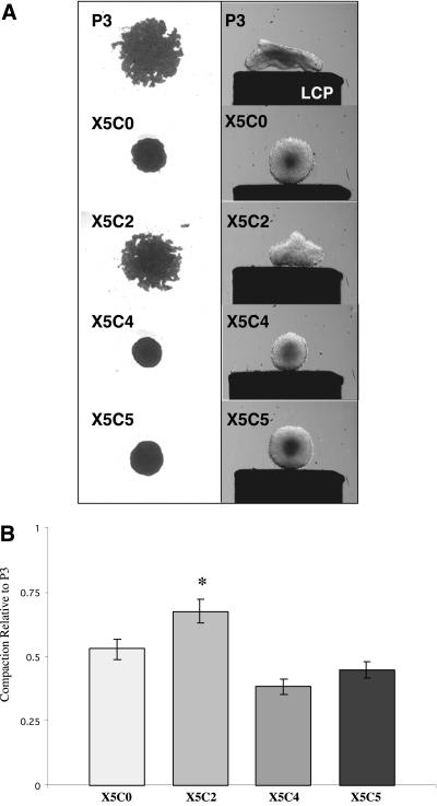
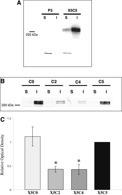
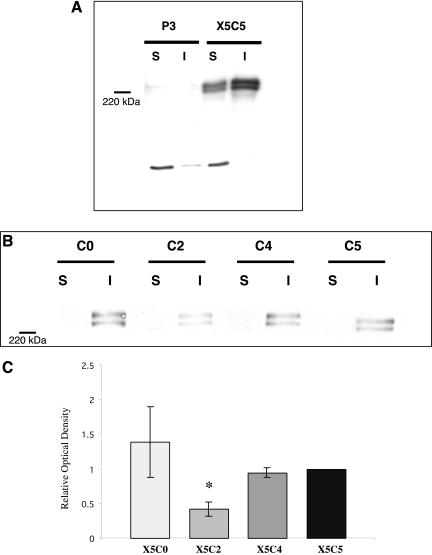
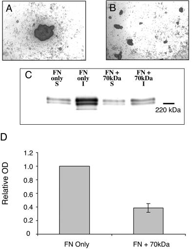
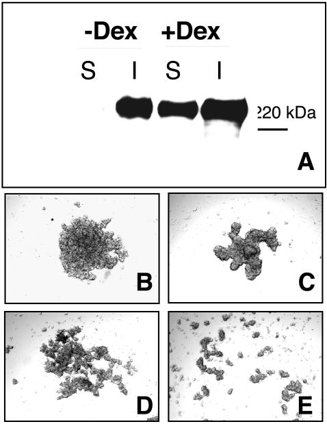
Integrin-extracellular matrix (ECM) interactions in two-dimensional (2D) culture systems are widely studied (Goldstein and DiMilla, 2002. J Biomed. Mater. Res. 59, 665-675; Koo et al., 2002. J. Cell Sci. 115, 1423-1433). Less understood is the role of the ECM in promoting intercellular cohesion in three-dimensional (3D) environments. We have demonstrated that the alpha5beta1-integrin mediates strong intercellular cohesion of 3D cellular aggregates (Robinson et al., 2003. J. Cell Sci. 116, 377-386). To further investigate the mechanism of alpha5beta1-mediated cohesivity, we used a series of chimeric alpha5beta1-integrin-expressing cells cultured as multilayer cellular aggregates. In these cell lines, the alpha5 subunit cytoplasmic domain distal to the GFFKR sequence was truncated, replaced with that of the integrin alpha4, the integrin alpha2, or maintained intact. Using these cells, alpha5beta1-integrin-mediated cell aggregation, compaction and cohesion were determined and correlated with FN matrix assembly. The data presented demonstrate that cells cultured in the absence of external mechanical support can assemble a FN matrix that promotes integrin-mediated aggregate compaction and cohesion. Further, inhibition of FN matrix assembly blocks the intercellular associations required for compaction, resulting in cell dispersal. These results demonstrate that FN matrix assembly contributes significantly to tissue cohesion and represents an alternative mechanism for regulating tissue architecture.


Figures

Figure 1.
Figure 1.
Determination of chimeric α5-integrin expression in transfected CHO B2 cells. (A) The X5C5, X5C4, X5C2, X5C0, and P3 cell lines were trypsinized from near confluent plates, washed and incubated with a mAb (mab) specific for the α5 extracellular domain, followed by incubation with a FITC-conjugated secondary antibody as described. The control panel (left) represents cells incubated with secondary antibody only. (B) Attached cells were lysed and total cellular lysates were analyzed by immunoblotting using a mab specific for the cytoplasmic (C) domain of the α5 or the α2 integrin. Alternately, lysates were incubated with a mab specific for the extracellular (X) domain of the α5 subunit. Immunocomplexes were recovered and analyzed by immunoblotting with a mab specific for the cytoplasmic domain of α4 or of α5, as indicated. In both cases, total lysates were analyzed by immunoblotting with a mab specific for the extracellular domain of the α5-Integrin to ensure equal loading.
Figure 2.
Figure 2.
Aggregate formation by chimeric integrin-expressing cells. The chimeric integrin-expressing cells (2.5 × 10⁶ cells/ml) were cultured in hanging drop assay for 2–3 d in FN-containing medium as described in MATERIALS AND METHODS. Aggregates were viewed using inverted bright field optics and images were captured using a spot camera connected to a MacIntosh G4 computer. Alternately, cell aggregates generated in hanging drop culture were transferred to shaker flasks and incubated in an orbital shaker for 2–3 d to allow for cell rearrangement and spheroid formation. Aggregate profiles are depicted on the lower compression plate (LCP) of the tensiometer as described in Robinson et al. (2003) (A). Pixel quantification of the aggregate images was performed using IPLabs imaging software and was expressed relative to the control P3 cells (B). Results are presented as the mean pixel value ± SEM of at least three separate experiments. *Statistical significance (p ≤ 0.05 vs. P3 cells) as determined by a one-way ANOVA followed by a Neuman-Keuls test.
Figure 3.
Figure 3.
FN matrix assembly in 2D culture does not correlate with aggregate compaction. The assembly of high-molecular-weight FN multimers by the P3, X5C5, X5C4, X5C2, and X5C0 cell lines was assessed using DOC differential solubilization assay. DOC-soluble and -insoluble fractions were separated and analyzed by immunoblotting. Matrix assembly of X5C5 cells was compared with P3 cells (A) and to the chimeric cells lines (B). Representative immunoblots are shown. Results for B were quantified by scanning densitometry using NIH Image software and expressed relative to the X5C5 cell line (C). A P3 signal was not detected and therefore, not included. The mean values ± SEM were calculated from at least three separate experiments. *Statistical significance (p ≤ 0.05 vs. X5C5 cells) as determined by a Student's unpaired t test.
Figure 4.
Figure 4.
FN matrix assembly in 3D culture correlates with aggregate compaction. P3, X5C5, X5C4, X5C2, and X5C0 cell lines were cultured as hanging drops for 16 h in the presence of 100 μg/ml exogenous rat plasma FN under tissue culture conditions. Aggregates were then harvested and the assembly of high-molecular-weight FN multimers was assessed as described in Figure 3. Matrix assembly of X5C5 cells in 3D culture was compared with P3 cells (A) and with the chimeric cells lines (B). Representative immunoblots are shown. Results for B were quantified by scanning densitometry using NIH Image software and expressed relative to the X5C5 cell line (C). A P3 signal was not detected and therefore, not included. The mean values ± SEM were calculated from at least three separate experiments. *Statistical significance (p ≤ 0.05 vs. X5C5 cells) as determined by a Student's unpaired t test.
Figure 5.
Figure 5.
X5C2 cells do not form fibrils in 3D culture. P3 (A), X5C5 (B), and X5C2 (C) cells were trypsinized from near-confluent plates and resuspended at 2.5 × 10⁶ cells/ml in FN-depleted medium (Corbett et al., 1997). Cells were incubated in the presence of 30 μg/ml rhodamine-labeled FN in 15-μl hanging drops for 24 h under tissue culture conditions. Hanging drops were then transferred to glass coverslips and visualized using inverted fluorescent optics. Bar, 20 μm.
Figure 6.
Figure 6.
Inhibition of FN matrix assembly by the 70-kDa FN fragment inhibits CHO-α5 aggregate formation. CHO-α5 cells (2.5 × 10⁶ cells/ml) were cultured in hanging drop assay in medium supplemented with 50 μg/ml FN, in the presence (B) or absence (A) of 25 μg/ml the 70-kDa amino-terminal FN fragment. Aggregates were viewed using inverted bright field optics. Alternately, hanging drops were harvested and the assembly of high-molecular-weight FN multimers assessed as described in Figure 3. (C) A representative immunoblot. In D, results were quantified by scanning densitometry using NIH Image software and expressed relative to the X5C5 cell line. The mean values ± SEM were calculated from at least three separate experiments. *Statistical significance (p ≤ 0.05 vs. X5C5 cells) as determined by a Student's unpaired t test.
Figure 7.
Figure 7.
The 70-kDa FN fragment inhibits HT1080 aggregate formation. HT1080 cells (2.5 × 10⁶ cells/ml) were cultured in hanging drop assay in the presence or absence of 10-7 M dexamethasone (A). Hanging drops were harvested and the assembly of high-molecular-weight FN multimers assessed as described in Figure 3. In separate experiments, HT1080 cells were cultured in FN-depleted medium (B and D) or medium supplemented with 50 μg/ml FN (C and E). The 70-kDa amino-terminal FN fragment (25 μg/ml) was added to inhibit matrix assembly (D and E). Aggregates were viewed using inverted bright field optics.

References

    1. Adamska, M., MacDonald, B.T., and Meisler, M.H. (2003). Doubleridge, a mouse mutant with defective compaction of the apical ectodermal ridge and normal dorsal-ventral patterning of the limb. Dev. Biol. 255, 350-362. - PubMed
    1. Akamatsu, H., Ichihara-Tanaka, K., Ozono, K., Kamiike, W., Matsuda, H., and Sekiguchi, K. (1996). Suppression of transformed phenotypes of human fibrosarcoma cells by overexpression of recombinant fibronectin. Cancer Res. 56, 4541-4546. - PubMed
    1. Akiyama, S.K., Aota, S., and Yamada, K.M. (1995). Function and receptor specificity of a minimal 20 kilodalton cell adhesive fragment of fibronectin. Cell Adhes. Commun. 3, 13-25. - PubMed
    1. Armstrong, P.B., and Armstrong, M.T. (2000). Intercellular invasion and the organizational stability of tissues: a role for fibronectin. Biochim. Biophys. Acta 1470, O9-O20. - PubMed
    1. Awad, H.A., Boivin, G.P., Dressler, M.R., Smith, F.N., Young, R.G., and Butler, D.L. (2003). Repair of patellar tendon injuries using a cell-collagen composite. J. Orthop. Res. 21, 420-431. - PubMed

LinkOut - more resources