. 2004 Mar;15(3):1334-46.
doi: 10.1091/mbc.e03-05-0296. Epub 2004 Jan 12.

A polycystin-1 multiprotein complex is disrupted in polycystic kidney disease cells

Affiliations

A polycystin-1 multiprotein complex is disrupted in polycystic kidney disease cells

Tamara Roitbak et al. Mol Biol Cell. 2004 Mar.

Abstract

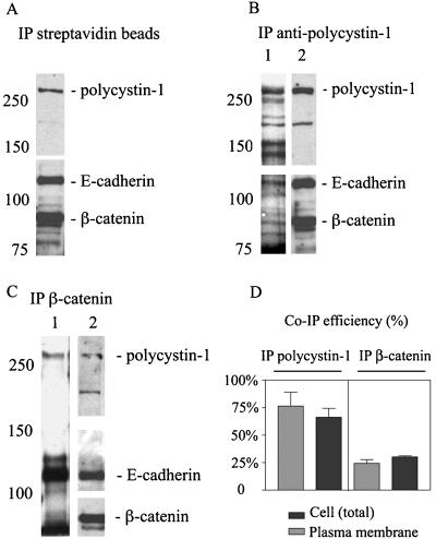
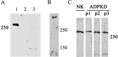
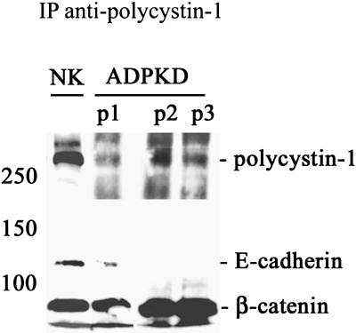
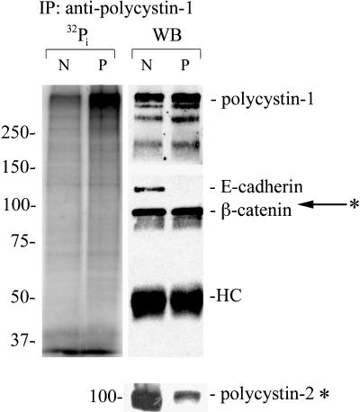
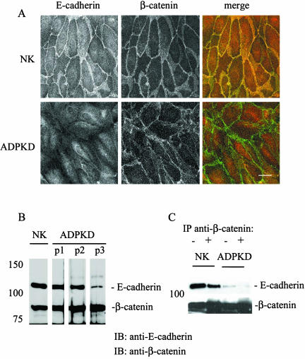
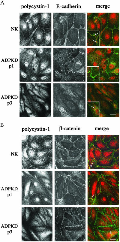
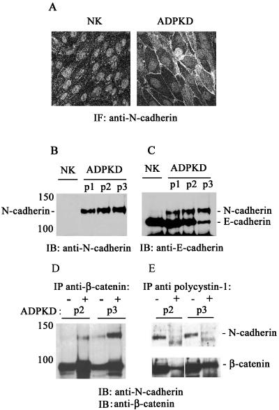
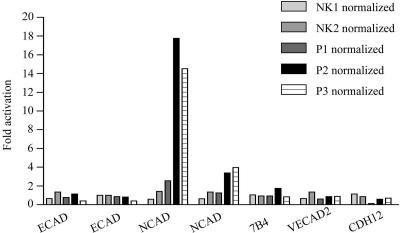
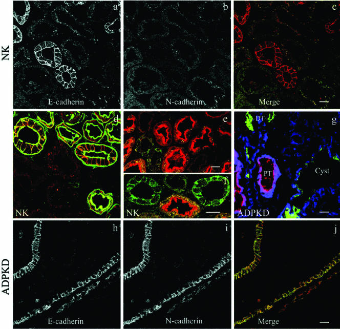
Autosomal dominant polycystic kidney disease (ADPKD) is typified by the accumulation of fluid-filled cysts and abnormalities in renal epithelial cell function. The disease is principally caused by mutations in the gene encoding polycystin-1, a large basolateral plasma membrane protein expressed in kidney epithelial cells. Our studies reveal that, in normal kidney cells, polycystin-1 forms a complex with the adherens junction protein E-cadherin and its associated catenins, suggesting a role in cell adhesion or polarity. In primary cells from ADPKD patients, the polycystin-1/polycystin-2/E-cadherin/beta-catenin complex was disrupted and both polycystin-1 and E-cadherin were depleted from the plasma membrane as a result of the increased phosphorylation of polycystin-1. The loss of E-cadherin was compensated by the transcriptional upregulation of the normally mesenchymal N-cadherin. Increased cell surface N-cadherin in the disease cells in turn stabilized the continued plasma membrane localization of beta-catenin in the absence of E-cadherin. The results suggest that enhanced phosphorylation of polycystin-1 in ADPKD cells precipitates changes in its localization and its ability to form protein complexes that are critical for the stabilization of adherens junctions and the maintenance of a fully differentiated polarized renal epithelium.


Figures

Figure 1.
Figure 1.
Polycystin-1 forms a complex with E-cadherin and β-catenin both at the cell membrane and in cytoplasm. Lysates from cell surface-biotinylated cells were precipitated with (A) streptavidin agarose, (B) antibodies against polycystin-1, or (C) antibodies against β-catenin. The proteins recovered in the immunoprecipitated fractions were probed with streptavidin-horseradish peroxidase to visualize the biotinylated plasma membrane proteins associated with polycystin-1 (B, lane 1) and β-catenin (C, lane 1). In parallel the streptavidin precipitates or the immunoprecipitates were also probed with the antibodies against polycystin-1, E-cadherin, and β-catenin to identify these proteins in the biotinylated samples (A, lane 2 in B and C). (D) The association of E-cadherin and polycystin-1 in multiprotein complexes was quantified and plotted using Prism software. Light bars represent quantification of the efficiency of coimmunoprecipitation of the two proteins in the cell surface complexes as shown in the lane 1 of the B and C. Dark bars represent quantification of the efficiency of coimmunoprecipitation of E-cadherin and polycystin-1 in the total cellular complexes as shown in the lane 2 of B and C. The values reflect the fraction of E-cadherin coprecipitated with polycystin-1 (lefthand bars) using the antibody against polycystin-1 or the fraction of polycystin-1 coprecipitated with E-cadherin (righthand bars) using and antibody against β-catenin for immunoprecipitation (mean, SE; n = 3). The ratio of E-cadherin coprecipitated with β-catenin was 97% and served to validate this approach.
Figure 2.
Figure 2.
Polycystin-1 expression levels are similar in normal kidney and ADPKD cells. (A, lanes 1-3) The specificity of our rabbit polyclonal antibody NM005 raised against a distal fragment of the polycystin-1 carboxy-terminus is demonstrated. Normal kidney cell lysates were immunoblotted with (1) NM005 antibody, directed against polycystin-1, (2) NM005 preincubated with antigen, and (3) preimmune serum. (B) The proteins from the normal kidney cell extract were separated on 4% polyacrylamide gel for 4 h, until only 150 and 250 MW prestained protein standard bands were left in the gel at the end of the electrophoresis. (C) Total cellular expression of polycystin-1 was evaluated in cell lysates prepared from the cells from normal kidney (NK) and three ADPKD patients (ADPKD P1, P2, and P3).
Figure 3.
Figure 3.
A normal multiprotein complex between polycystin-1, E-cadherin, and β-catenin is disrupted in ADPKD cells. Cell lysates prepared from normal kidney (NK) and three ADPKD patients (ADPKD P1, P2, and P3) were immunoprecipitated (IP) with our antipolycystin antibody under nondenaturing conditions. Immunoprecipitates were immunoblotted with antibodies directed against polycystin-1, E-cadherin, and β-catenin to determine expression of these proteins in the immunoprecipitates.
Figure 4.
Figure 4.
Polycystin-1 is highly phosphorylated in ADPKD cells. The [32P]orthophosphate labeled samples (two left lanes) and unlabeled samples (right lanes) from normal kidney (N) and ADPKD (P) cells were immunoprecipitated (IP) with antipolycystin antibody under nondenaturing conditions. Immunoprecipitates were immunoblotted with antibodies directed against polycystin-1, polycystin-2, E-cadherin, and β-catenin to confirm their presence or absence in the immunoprecipitates and establish the identity of the 32P-labeled proteins. The relative migration of polycystin-2 is indicated with an arrow. Polycystin-2 was not significantly phosphorylated. Because polycystin-2 has a molecular weight just above beta-catenin this immunoblot is shown separately. HC, IgG heavy chain.
Figure 5.
Figure 5.
E-cadherin is not expressed at the lateral plasma membrane and does not form a complex with β-catenin in ADPKD cells. (A) Confluent monolayers of primary normal kidney (NK) and ADPKD (patient 2) were immunostained for E-cadherin (red) and β-catenin (green). Samples were imaged on a Zeiss LSM510 (Thornwood, NY), and 0.5-μm sections are shown of each of the individual protein distributions (black and white images) as well as a merge (color) to show any colocalization in yellow. Bar, 20 μm. (B) Total cellular expression levels of E-cadherin and β-catenin were evaluated by immunoblotting samples from normal kidney (NK) and three ADPKD patients (ADPKD P1, P2, and P3). (C) The association of E-cadherin and β-catenin was monitored in confluent normal kidney (NK) and ADPKD cells by coimmunoprecipitation studies. Cell lysates prepared under nondenaturing conditions were immunoprecipitated with a specific mAb directed against β-catenin. Cell lysates (-) and immunoprecipitates (+) were immunoblotted (IB) with antibodies directed against E-cadherin and β-catenin.
Figure 6.
Figure 6.
Comparison of polycystin-1, E-cadherin, and β-catenin localizations in normal kidney and ADPKD cells. Confluent normal kidney (NK) and ADPKD cells were double-labeled with polyclonal NM005 antibody directed against polycystin-1 and monoclonal antibodies directed against (A) E-cadherin and (B) β-catenin and imaged as in Figure 5. Shown are one NK sample and two ADPKD patient samples (P1 and P3). Black and white panels show individual localizations of polycystin-1, E-cadherin, and β-catenin as indicated. Dual staining is presented in the merged images where polycystin-1 is in the red channel, and (A) E-cadherin and (B) β-catenin are in the green channel. Colocalization sites of (A) polycystin-1 and E-cadherin and (B) polycystin-1 and β-catenin are seen as a yellow overlap pattern. Insets in the merged images (A) show a twofold enlarged detail of sites where the two proteins are colocalized. Bar, 20 μm.
Figure 7.
Figure 7.
N-cadherin replaces E-cadherin at the plasma membrane of ADPKD cells and forms a complex with β-catenin. (A) N-cadherin was visualized by immunofluorescence staining of normal kidney (NK) and ADPKD cells using a mouse mAb directed against N-cadherin. Bar, 20 μm. (B) The total cellular expression of N-cadherin was analyzed in normal kidney cell lysates (NK) and cell lysates from three ADPKD patients (ADPKD P1, P2, and P3) by immunoblotting (IB). (C) The same blot was subsequently probed with an antibody directed against E-cadherin to reveal the total levels of E-cadherin expression in the same samples. (D and E) The presence of N-cadherin/β-catenin complexes and their association with polycystin-1 were assessed by coimmunoprecipitation experiments. Cell lysates from two ADPKD patients (ADPKD P2, P3) were immunoprecipitated (IP) with an antibody directed against (D) β-catenin and (E) against polycystin-1. The cell lysates (-) and immunoprecipitates (+) were immunoblotted (IB) with a polyclonal antibody directed against N-cadherin and subsequently immunoblotted with an antibody directed against β-catenin.
Figure 8.
Figure 8.
The level of N-cadherin RNA expression is significantly elevated in ADPKD cells. Total RNA was prepared from the cultured primary epithelial cells from two different normal kidney (NK1 and NK2) and three ADPKD samples (P1, P2, and P3) and used for hybridization to Affymetrix Gene Chips. The data were normalized against the median value of the normal kidney samples. The fold RNA expression values were plotted using Prism Software. Plotted are E-cadherin (ECAD), N-cadherin (NCAD), vascular epithelial cadherin (7B4), vascular epithelial cadherin 2 or protocadherin (VECAD2), and neuronal N-cadherin precursor protein (CDH12). The hybridization to two different E- and N-cadherin oligonucleotides on the chips is graphed separately.
Figure 9.
Figure 9.
ADPKD cysts express both N- and E-cadherin. Tissue sections of (a-f) normal kidney cortex and (g-j) ADPKD cystic tissue were double- or triple-labeled with (a, c, d-f, h, and j) polyclonal antibodies directed against E-cadherin, (b, c, g, i, and j) a mAb against N-cadherin, and (d-g) tubule-specific lectins. (a-c) E-cadherin (red) and N-cadherin (green) are expressed in distinct normal kidney tubules. (d) E-cadherin (red) is colocalized with peanut lectin (green) specific for distal parts of the nephron (distal tubules and collecting duct). (e and f) E-cadherin (green) exhibits a distinct distribution from asparagus lectin (red), a marker for the proximal tubules. (g) N-cadherin is overexpressed in early cysts. ADPKD tissue triple-labeled with an mAb directed against N-cadherin (blue), with lectins for proximal tubules (PT, red) and distal tubules (DT, green). (h-j) E-cadherin (red) and N-cadherin (green) exhibit overlapping distributions in ADPKD cysts. Bars, 20 μm.

Similar articles

Cited by

References

    1. Angst, B.D., Marcozzi, C., and Magee, A.I. (2001). The cadherin superfamily: diversity in form and function. J. Cell Sci. 114, 629-641. - PubMed
    1. Arnaout, M.A. (2001). Molecular genetics and pathogenesis of autosomal dominant polycystic kidney disease. Annu. Rev. Med. 52, 93-123. - PubMed
    1. Arnould, T., Kim, E., Tsiokas, L., Jochimsen, F., Gruning, W., Chang, J.D., and Walz, G. (1998). The polycystic kidney disease 1 gene product mediates protein kinase C alpha-dependent and c-Jun N-terminal kinase-dependent activation of the transcription factor AP-1. J. Biol. Chem. 273, 6013-6018. - PubMed
    1. Balsamo, J., and Lilien, J. (1990). N-cadherin is stably associated with and is an acceptor for a cell surface N-acetylgalactosaminylphosphotransferase. J. Biol. Chem. 265, 2923-2928. - PubMed
    1. Barbar, E., Rola-Pleszczynski, M., Payet, M.D., and Dupuis, G. (2003). Protein kinase C inhibits the transplasma membrane influx of Ca2+ triggered by 4-aminopyridine in Jurkat T lymphocytes. Biochim. Biophys. Acta 1622, 89-98. - PubMed

Publication types