. 2004 Feb;13(2):381-90.
doi: 10.1110/ps.03336804. Epub 2004 Jan 10.

The folding landscape of Streptomyces griseus protease B reveals the energetic costs and benefits associated with evolving kinetic stability

Affiliations

The folding landscape of Streptomyces griseus protease B reveals the energetic costs and benefits associated with evolving kinetic stability

Stephanie M E Truhlar et al. Protein Sci. 2004 Feb.

Abstract

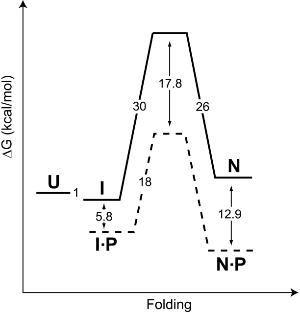
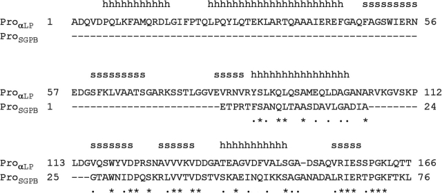
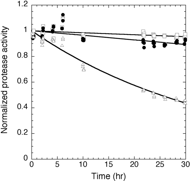
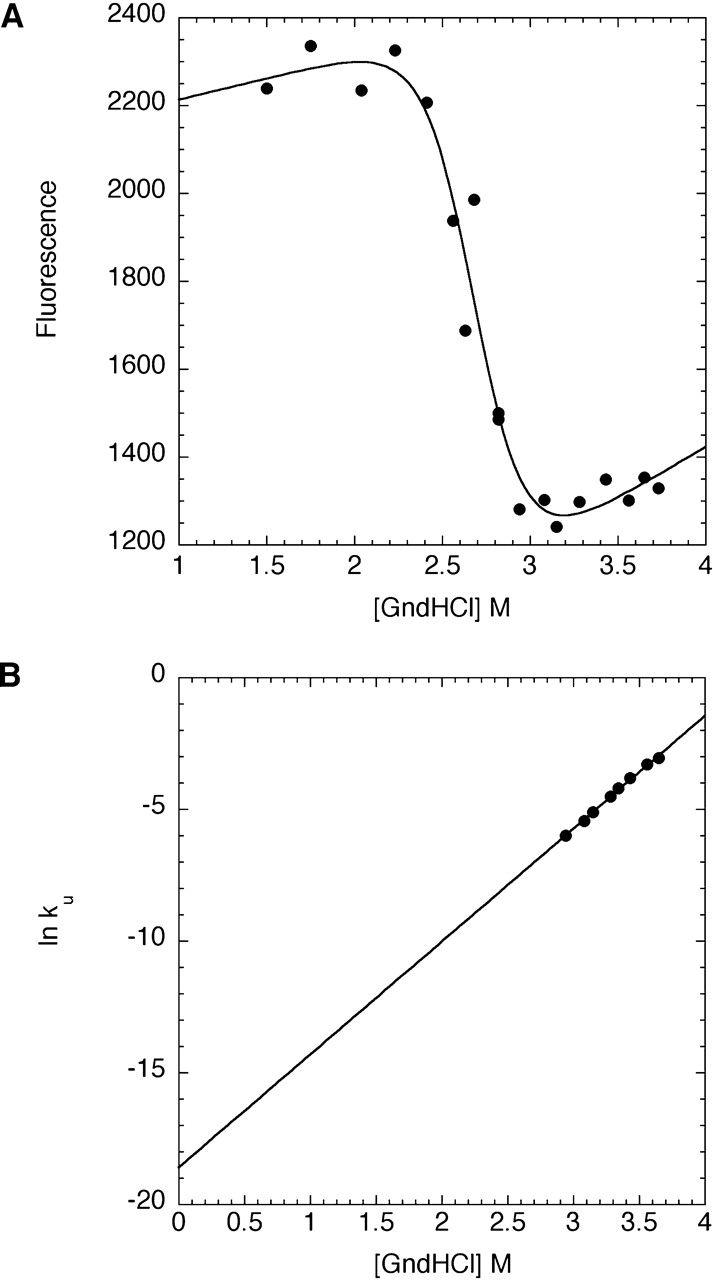
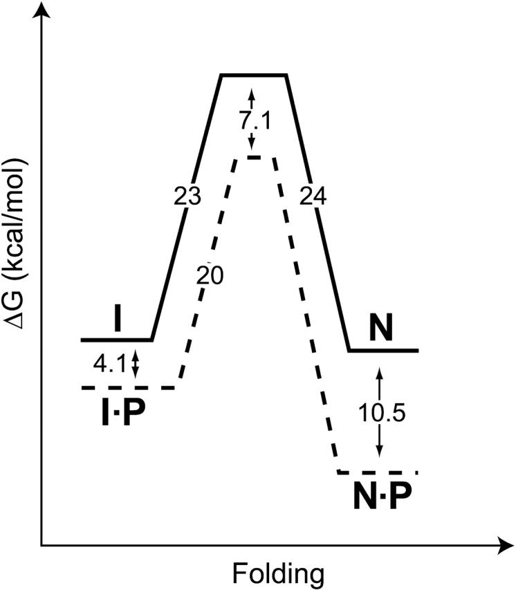
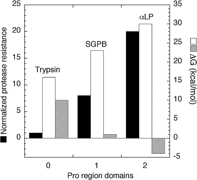
Like most extracellular bacterial proteases, Streptomyces griseus protease B (SGPB) and alpha-lytic protease (alphaLP) are synthesized with covalently attached pro regions necessary for their folding. In this article, we characterize the folding free energy landscape of SGPB and compare it to the folding landscapes of alphaLP and trypsin, a mammalian homolog that folds independently of its zymogen peptide. In contrast to the thermodynamically stable native state of trypsin, SGPB and alphaLP fold to native states that are thermodynamically marginally stable or unstable, respectively. Instead, their apparent stability arises kinetically, from unfolding free energy barriers that are both large and highly cooperative. The unique unfolding transitions of SGPB and alphaLP extend their functional lifetimes under highly degradatory conditions beyond that seen for trypsin; however, the penalty for evolving kinetic stability is remarkably large in that each factor of 2.4-8 in protease resistance is accompanied by a cost of ~10(5) in the spontaneous folding rate and ~5-9 kcal/mole in thermodynamic stability. These penalties have been overcome by the coevolution of increasingly effective pro regions to facilitate folding. Despite these costs, kinetic stability appears to be a potent mechanism for developing native-state properties that maximize protease longevity.


Figures

Figure 1.
Figure 1.
The folding free energy landscape for αLP at 0°C (modified from Peters et al. 1998; Sohl et al. 1998; Derman and Agard 2000; S. Jaswal, unpubl.). Solid lines and dashed lines depict the free energy barriers encountered by αLP in the absence and presence of its pro region (P), respectively. The αLP native state (N) is thermodynamically less stable than either the intermediate state (I) or the fully unfolded molecule (U). Free energies of activation were calculated from folding and unfolding rates using the transition state theory (Glasstone et al. 1941). The stability of the αLP intermediate state was measured at 4°C, and pro region binding to the native state was measured at 25°C.
Figure 2.
Figure 2.
Sequence alignment of the αLP and SGPB pro regions. Homology models of ProSGPB show that it is compatible with the α-helical (h) and β-strand (s) ProαLP structure (Sauter et al. 1998) indicated above its sequence. Pro region sequence identity (*) and similarity (•) are indicated below the sequences.
Figure 3.
Figure 3.
Characterization of ProSGPB by circular dichroism (CD) and inhibition of SGPB. (A) The molar ellipticity at 220 nm of ProSGPB as a function of ethylene glycol concentration was fit as described (see Materials and Methods) to determine the thermodynamic stability of ProSGPBGf = 2.4 ± 0.3 kcal/mole). (B) The CD spectrum, plotted as molar ellipticity versus wavelength, of SGPB (open circles) displays primarily β-sheet secondary structure, whereas the spectrum of ProSGPB (open triangles) appears to be almost completely random coil. The spectrum of the noninteracting proteins (filled squares) is significantly different from the spectrum of the mixed proteins (filled circles), which displays increased α-helical signal, consistent with the formation of the predicted helical structure of ProSGPB, upon binding to SGPB (B, inset). Every fifth data point is shown to simplify the spectra. (C) SGPB proteolytic activity was measured in the presence of increasing amounts of ProSGPB, and the data were fit to the competitive inhibitor equation to determine the affinity of the interaction between ProSGPB and the SGPB native state (Ki = 19 ± 2 nM).
Figure 4.
Figure 4.
Kinetic analysis of SGPB folding and unfolding. (A) The concentration dependence of the ProSGPB-catalyzed refolding of SGPB shows Michaelis-Menten type behavior (kcat = 0.0010 ± 0.0001 sec−1, KM = 530 ± 66 μM). (B) The rate of spontaneous SGPB folding (kf = 2.7 × 10⁻⁶ ± 0.1 × 10⁻⁶ sec−1) was calculated from a linear fit of the fraction of folded SGPB as a function of refolding time. (C) The rate of SGPB unfolding in the absence of denaturant (GndHCl) was calculated from a linear fit of the denaturant dependence of the unfolding rates (ku = 7.4 × 10⁻⁷ ± 0.4 × 10⁻⁷ sec−1).
Figure 5.
Figure 5.
Protease resistance of αLP, SGPB, and trypsin. Coincubation of αLP, SGPB, and trypsin under strongly proteolytic conditions at 37°C reveals that SGPB (filled circles) is inactivated 2.4 times faster than αLP (open squares) (kinact, SGPB = 1.0 × 10⁻⁶ ± 0.2 × 10⁻⁶ sec−1, kinact,αLP = 4.2 × 10⁻⁷ ± 1.5 × 10⁻⁷ sec−1). SGPB (data not shown) and αLP (S. Jaswal, unpubl.) are inactivated at approximately the same rate as their global unfolding at this temperature. Both SGPB and αLP significantly outlast trypsin (open triangles) (kinact, trypsin = 7.8 × 10⁻⁶ ± 0.2 × 10⁻⁶ sec−1).
Figure 6.
Figure 6.
Equilibrium and kinetic analysis of trypsin unfolding. (A) The equilibrium fluorescence of trypsin as a function of denaturant was fit to the linear free energy model to determine the thermodynamic stability of trypsin (ΔGu = 10 ± 2 kcal/mole). (B) Each symbol denotes the logarithm of the rate constant for trypsin unfolding at a given denaturant (GndHCl) concentration. Linear extrapolation of the data yields the unfolding rate in the absence of GndHCl (ku = 8.4 × 10⁻⁹ ± 0.4 × 10⁻⁹ sec−1).
Figure 7.
Figure 7.
SGPB folding free energy landscape at 0°C. Solid lines depict the free energies of activation for SGPB folding and unfolding (from Fig. 4B,C ▶). Dashed lines illustrate the effect of ProSGPB (P) on the free energy profile (from Figs. 3C ▶, 4A ▶). Unlike αLP, the SGPB native state (N) is marginally stable compared to its intermediate state (I). Free energies of activation were calculated from folding and unfolding rates using transition state theory (Glasstone et al. 1941). Pro region binding to the native state was measured at 25°C.
Figure 8.
Figure 8.
Comparative analysis of protease resistance and the folding energetics of trypsin, SGPB, and αLP. By arranging the three proteases in order of their proteolytic sensitivity we can compare the consequences of evolving increased protease resistance (black bars, normalized protease resistance). A dramatically increased barrier to folding (white bars, kcal/mole) and a loss of thermodynamic stability (striped bars, kcal/mole) seem directly correlated with an increase in proteolytic resistance. Circumventing these difficulties requires increasingly complex pro regions. Trypsin folds without a pro region, whereas SGPB requires a single-domain pro region and αLP requires a two-domain pro region to facilitate folding.

References

    1. Anderson, D.E., Peters, R.J., Wilk, B., and Agard, D. 1999. α-Lytic protease precursor: Characterization of a structured folding intermediate. Biochemistry 38 4728–4735. - PubMed
    1. Baardsnes, J., Sidhu, S., MacLeod, A., Elliott, J., Morden, D., Watson, J., and Borgford, T. 1998. Streptomyces griseus protease B: Secretion correlates with the length of the propeptide. J. Bacteriol. 180 3241–3244. - PMC - PubMed
    1. Baker, D., Sohl, J.L., and Agard, D.A. 1992. A protein-folding reaction under kinetic control. Nature 356 263–265. - PubMed
    1. Bhattacharjya, S., Xu, P., and Ni, F. 2000. Sequence-specific 1H, 15N and 13C resonance assignments of the inhibitory prodomain of human furin. J. Biomol. NMR 16 275–276. - PubMed
    1. Bryan, P., Wang, L., Hoskins, J., Ruvinov, S., Strausberg, S., Alexander, P., Almog, O., Gilliland, G., and Gallagher, T. 1995. Catalysis of a protein folding reaction: Mechanistic implications of the 2.0 Å structure of the subtilisin–prodomain complex. Biochemistry 34 10310–10318. - PubMed

Publication types

LinkOut - more resources