. 2004 Jan 20;101(3):757-62.
doi: 10.1073/pnas.2036345100. Epub 2004 Jan 12.

MLL 5 protein forms intranuclear foci, and overexpression inhibits cell cycle progression

Affiliations

MLL 5 protein forms intranuclear foci, and overexpression inhibits cell cycle progression

Lih-Wen Deng et al. Proc Natl Acad Sci U S A. .

Abstract

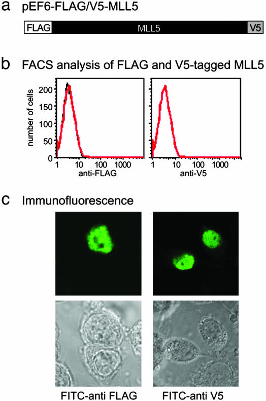
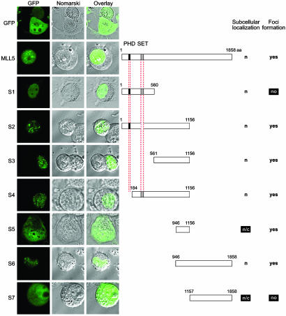
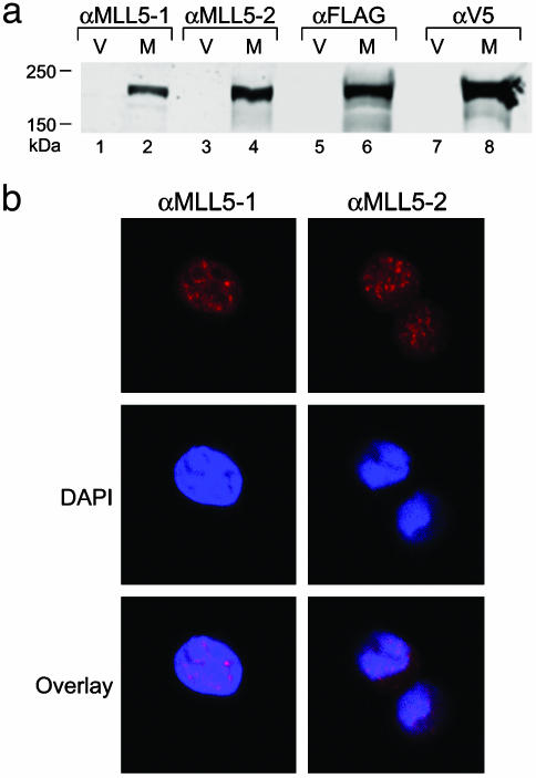
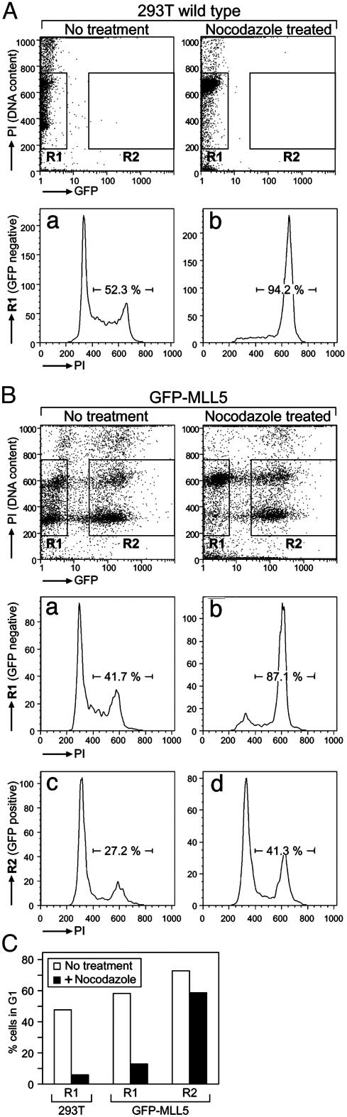
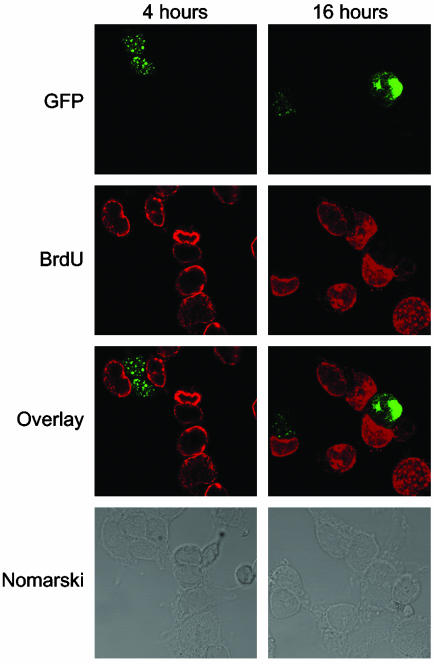
MLL5 is a mammalian trithorax group (trx-G) gene identified within chromosome band 7q22, a frequently deleted element found in cytogenetic aberrations of acute myeloid malignancies. MLL5 cDNA was linked with the FLAG and V5 tags at the N and C terminus, respectively, and transfected into 293T cells. Immunofluoresence staining of the expressed tagged MLL5 protein showed localization to the nucleus and exclusion from nucleoli, and no surface staining was detected. Both ectopically introduced and endogenous MLL5 protein displayed a speckled nuclear distribution. By using a series of MLL5-truncated mutants fused with enhanced GFP, a domain (residues 945-1,156) required for foci accumulation was identified, and regions containing functional nuclear localization signals were mapped. Ectopic overexpression of GFP-MLL5 induced cell cycle arrest in G(1) phase. This inhibition of cell cycle progression was indicated by delayed progression into nocodazole-induced mitotic arrest and was confirmed by a lack of BrdUrd incorporation. These findings suggest that MLL5 forms intranuclear protein complexes that may play an important role in chromatin remodeling and cellular growth suppression.


Figures

Fig. 1.
Fig. 1.
Nuclear localization of MLL5 protein. (a) Full-length MLL5 was tagged with FLAG and V5 epitopes at the N and C termini, respectively, and cloned into the pEF6 vector. (b) The 293T cells were transiently transfected with the pEF6-flag/V5-MLL5 vector for 48 h, stained with 10 μg/ml FITC-conjugated anti-FLAG or FITC-conjugated anti-V5 mAbs for 1 h, and analyzed by flow cytometry (red). Staining for isotype controls is in black. (c) pEF6-flag/v5-MLL5 transfectants were fixed in prechilled 100% methanol for 10 min at –20°C, rehydrated in PBS, and permeabilized in 0.1% Triton X-100/PBS for 5 min. Cells were blocked in PBS containing 3% BSA at RT for 30 min followed by incubation for 1 h at RT with FITC-conjugated anti-FLAG and anti-V5 mAbs.
Fig. 2.
Fig. 2.
Mapping of MLL5 elements directing nuclear localization and foci formation. Full-length and segments of MLL5 protein were cloned into the pEGFPC1 vector with enhanced GFP fused to their N termini. These constructs were transiently transfected into 293T cells, and live cells were imaged by confocal microscopy. Fluorescence micrographs are shown on the left, with GFP fluorescence followed by Nomarski images and by overlays of Nomarski and fluorescence images. Schematics of the corresponding constructs are displayed on the right. The PHD zinc finger is colored in black, and the SET domain is colored in gray. Amino acid numbers are denoted above each construct. Observed subcellular localization is indicated as nuclear (n) or cytoplasmic (c). The ability to form intranuclear foci is indicated on the right.
Fig. 3.
Fig. 3.
(a) Western blot analysis of FLAG-V5-tagged MLL5 expression. Lanes contained whole-cell lysates of 293T cells transfected with either pEF6 vector alone (V) or pEF6-flag-V5-MLL5 (M). Blots were probed with Abs αMLL5-1, αMLL5-2, anti-FLAG mAb, or anti-V5 mAb. The polyclonal Abs αMLL5-1 and αMLL5-2 were raised against synthetic peptides corresponding to amino acids 227–241 and 801–815 of the MLL5 protein, respectively. (b) Endogenous immunofluorescent staining of MLL5 in 293T cells by using rabbit polyclonal Abs αMLL5-1 or αMLL5-2, followed by Alexa 568-conjugated anti-rabbit polyclonal Abs (red). Nuclei were counterstained with 4′,6-diamidino-2′-phenylindole dihydrochloride (DAPI; blue).
Fig. 4.
Fig. 4.
Induction of G1 cell cycle arrest by GFP-MLL5 expression. WT 293T cells or 293T cells transfected with GFP-MLL5 were treated with or without nocodazole for 16 h and then fixed, permeabilized, and stained with PI as described in Methods for cell cycle analysis. (A) WT 293T cells displayed a shift in population to cells with 4N DNA content (G2/M phase) after nocodazole treatment (a and b). (Upper) PI staining (DNA content) was plotted against GFP fluorescence with GFP-negative cells gated on as R1 and positive transfectants gated on as R2. (Lower) DNA content histograms for these respective gates are displayed. (B) The untransfected population (R1) of the GFP-MLL5 transfectant cell culture also displayed a shift to 4N DNA content after nocodazole arrest (a and b). In GFP-MLL5-positive transfectants (R2), however, the profile of cells with 4N content remained constant even after 16-h treatment with nocodazole (c and d). (C) A quantitation of the percentage of cells in G1 phase of the above populations with or without nocodazole treatment indicates an arrest of GFP-MLL5-positive cells (R2) in G1 phase.
Fig. 5.
Fig. 5.
Overexpression of MLL5 protein in 293T cells inhibits DNA replication. After transfection (48 h), GFP-MLL5 cells were incubated with 20 μg/ml BrdUrd for 4 or 16 h. Resulting cultures were fixed, permeabilized, treated with 1 N HCl to expose BrdUrd sites, and stained with Alexa 568-conjugated anti-BrdUrd mAb (red) and Alexa 488-conjugated anti-GFP mAb (green). The third panel displays an overlay of BrdUrd and GFP-MLL5 staining, and the fourth panel shows the corresponding Nomarski images.

References

    1. Ziemin-van der Poel, S., McCabe, N. R., Gill, H. J., Espinosa, R., III, Patel, Y., Harden, A., Rubinelli, P., Smith, S. D., LeBeau, M. M. & Rowley, J. D. (1991) Proc. Natl. Acad. Sci. USA 88, 10735–10739. - PMC - PubMed
    1. Gu, Y., Nakamura, T., Alder, H., Prasad, R., Canaani, O., Cimino, G., Croce, C. M. & Canaani, E. (1992) Cell 71, 701–708. - PubMed
    1. Tkachuk, D. C., Kohler, S. & Cleary, M. L. (1992) Cell 71, 691–700. - PubMed
    1. Djabali, M., Selleri, L., Parry, P., Bower, M., Young, B. D. & Evans, G. A. (1992) Nat. Genet. 2, 113–118. - PubMed
    1. Dimartino, J. F. & Cleary, M. L. (1999) Br. J. Haematol. 106, 614–626. - PubMed

Publication types

MeSH terms

LinkOut - more resources