. 2004 Jan 20;101(3):817-22.
doi: 10.1073/pnas.0305555101. Epub 2004 Jan 12.

Ectopic expression of the homeobox gene Cdx2 is the transforming event in a mouse model of t(12;13)(p13;q12) acute myeloid leukemia

Affiliations

Ectopic expression of the homeobox gene Cdx2 is the transforming event in a mouse model of t(12;13)(p13;q12) acute myeloid leukemia

Vijay P S Rawat et al. Proc Natl Acad Sci U S A. .

Abstract

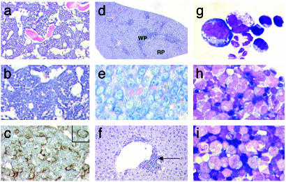
Creation of fusion genes by balanced chromosomal translocations is one of the hallmarks of acute myeloid leukemia (AML) and is considered one of the key leukemogenic events in this disease. In t(12;13)(p13;q12) AML, ectopic expression of the homeobox gene CDX2 was detected in addition to expression of the ETV6-CDX2 fusion gene, generated by the chromosomal translocation. Here we show in a murine model of t(12;13)(p13;q12) AML that myeloid leukemogenesis is induced by the ectopic expression of CDX2 and not by the ETV6-CDX2 chimeric gene. Mice transplanted with bone marrow cells retrovirally engineered to express Cdx2 rapidly succumbed to fatal and transplantable AML. The transforming capacity of Cdx2 depended on an intact homeodomain and the N-terminal transactivation domain. Transplantation of bone marrow cells expressing ETV6-CDX2 failed to induce leukemia. Furthermore, coexpression of ETV6-CDX2 and Cdx2 in bone marrow cells did not accelerate the course of disease in transplanted mice compared to Cdx2 alone. These data demonstrate that activation of a protooncogene by a balanced chromosomal translocation can be the pivotal leukemogenic event in AML, characterized by the expression of a leukemia-specific fusion gene. Furthermore, these findings link protooncogene activation to myeloid leukemogenesis, an oncogenic mechanism so far associated mainly with lymphoid leukemias and lymphomas.


Figures

Fig. 1.
Fig. 1.
(a) Retroviral vectors used to express ETV6-CDX2, Cdx2, and the different Cdx2 mutants in murine BM. IRES, internal ribosomal entry site. (b) Western blot analysis of cellular extracts from NIH 3T3 or E86 cells transfected with the different constructs. The molecular mass is indicated.
Fig. 2.
Fig. 2.
Survival curve of mice transplanted with BM cells expressing Cdx2 (n = 18), ETV6-CDX2 (n = 9), or coexpressing Cdx2 and the fusion gene (n = 13). The control group was injected with BM infected with the GFP empty retrovirus (n = 7). The survival time of secondary recipient mice, transplanted with BM from diseased primary Cdx2 or ETV6-CDX2 and Cdx2 recipients, is indicated.
Fig. 3.
Fig. 3.
Histological analysis of diseased Cdx2 mice. (a) BM [hematoxylin/eosin (H&E)]. Immunohistochemistry of the BM (×200) for N-acetyl-chloroacetate esterase (×400) (b) and CD34 expression (×640) (c). Histology of the spleen H&E (×25) (d) and Giemsa staining (×640) (e) and liver with perivascular infiltration (×200) (f). Cytospin preparations from PB (g), BM (h), and spleen (i) (all ×1,000).
Fig. 4.
Fig. 4.
(a) Flow cytometry from a representative leukemic Cdx2 mouse from PB, BM, and spleen in comparison to a GFP control animal. Cells were stained for the myeloid markers Gr1 and Mac1 and the lymphoid marker B220. The proportion of positive cells within the GFP+ compartment is indicated. (b) Southern blot analyses of genomic DNA from BM, PB, and spleens of representative leukemic Cdx2 mice. Genomic DNA was digested with EcoRI, which cuts once in the provirus, to determine the number of provirus integrants. Signals with different intensity, indicating the presence of different leukemic clones, are indicated. Full-length provirus integration was documented by digestion with NheI, which cuts only in the LTRs of the provirus.
Fig. 5.
Fig. 5.
(a) Total number of d12 CFU-S colonies derived per culture initiated with 1 × 10⁵ cells transduced with the different viruses after 1 wk in liquid culture. The median is indicated. (b) Expression of Meis1 and Hoxa9 analyzed by RT-PCR in Scalin+ BM cells isolated from a Cdx2 mouse or a control mouse. The number of PCR cycles for each gene was chosen to stop the reaction in the linear phase of the amplification (25 cycles for mβ-2 microglobulin, 35 cycles for Meis1 and Hoxa9). C, control; Cdx, Cdx2.

Similar articles

Cited by

References

    1. Rowley, J. D. (1999) Semin. Hematol. 36, 59–72. - PubMed
    1. Bohlander, S. K. (2000) Cytogenet. Cell Genet. 91, 52–56. - PubMed
    1. Look, A. T. (1997) Science 278, 1059–1064. - PubMed
    1. Pineault, N., Buske, C., Feuring-Buske, M., Abramovich, C., Rosten, P., Hogge, D. E., Aplan, P. D. & Humphries, R. K. (2003) Blood 101, 4529–4538. - PubMed
    1. Kroon, E., Krosl, J., Thorsteinsdottir, U., Baban, S., Buchberg, A. M. & Sauvageau, G. (1998) EMBO J. 17, 3714–3725. - PMC - PubMed

Publication types

MeSH terms