Review
. 2003:62:19-111.
doi: 10.1016/s0065-3527(03)62002-6.

Evolution of cell recognition by viruses: a source of biological novelty with medical implications

Affiliations
Review

Evolution of cell recognition by viruses: a source of biological novelty with medical implications

Eric Baranowski et al. Adv Virus Res. 2003.

Abstract

The picture beginning to form from genome analyses of viruses, unicellular organisms, and multicellular organisms is that viruses have shared functional modules with cells. A process of coevolution has probably involved exchanges of genetic information between cells and viruses for long evolutionary periods. From this point of view present-day viruses show flexibility in receptor usage and a capacity to alter through mutation their receptor recognition specificity. It is possible that for the complex DNA viruses, due to a likely limited tolerance to generalized high mutation rates, modifications in receptor specificity will be less frequent than for RNA viruses, albeit with similar biological consequences once they occur. It is found that different receptors, or allelic forms of one receptor, may be used with different efficiency and receptor affinities are probably modified by mutation and selection. Receptor abundance and its affinity for a virus may modulate not only the efficiency of infection, but also the capacity of the virus to diffuse toward other sites of the organism. The chapter concludes that receptors may be shared by different, unrelated viruses and that one virus may use several receptors and may expand its receptor specificity in ways that, at present, are largely unpredictable.


Figures
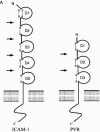
Fig 1
Fig 1
(A) Representation and comparison of the domain structure of ICAM-1 (CD54), the receptor for the major group rhinoviruses, and the poliovirus receptor (PVR or CD155). The immunoglobulin-like domains (labeled D1–D5 or D1–D3) are represented schematically by a circle closed by one or two disulfide bonds. The different Ig domains are linked by a flexible peptide chain. Hinge points are indicated by arrows. (B) Cryo-EM reconstruction showing the complex of HRV16 with its ICAM-1 receptor (from Kolatkar et al., 1999) virus is represented as a gray-scale surface. D1 and D2 domains of ICAM-1 are colored red. (C) Cryo-EM reconstruction of the complex of PV1(M) (gray) with PVR (yellow) (from Xing et al., 2000). (D) ICAM-1 and PVR-binding modes. Stereoview of the ICAM-1 (red) docked onto one icosahedral asymmetric unit of HRV16 (gray) using the cryo-EM map as a guide (PDB accession code 1D3E). The structure of PVR in complex with PV1 (M) (PDB accession code 1DGI) was superimposed for comparison (yellow). ICAM-1 contacts primarily the floor and south wall of the HRV16 canyon. In contrast, PVR overlaps the north and south walls, as well as the floor of the canyon, making additional contacts with the viral surface.
Fig 1
Fig 1
(A) Representation and comparison of the domain structure of ICAM-1 (CD54), the receptor for the major group rhinoviruses, and the poliovirus receptor (PVR or CD155). The immunoglobulin-like domains (labeled D1–D5 or D1–D3) are represented schematically by a circle closed by one or two disulfide bonds. The different Ig domains are linked by a flexible peptide chain. Hinge points are indicated by arrows. (B) Cryo-EM reconstruction showing the complex of HRV16 with its ICAM-1 receptor (from Kolatkar et al., 1999) virus is represented as a gray-scale surface. D1 and D2 domains of ICAM-1 are colored red. (C) Cryo-EM reconstruction of the complex of PV1(M) (gray) with PVR (yellow) (from Xing et al., 2000). (D) ICAM-1 and PVR-binding modes. Stereoview of the ICAM-1 (red) docked onto one icosahedral asymmetric unit of HRV16 (gray) using the cryo-EM map as a guide (PDB accession code 1D3E). The structure of PVR in complex with PV1 (M) (PDB accession code 1DGI) was superimposed for comparison (yellow). ICAM-1 contacts primarily the floor and south wall of the HRV16 canyon. In contrast, PVR overlaps the north and south walls, as well as the floor of the canyon, making additional contacts with the viral surface.
Fig 2
Fig 2
(A) Ribbon representation of a CS8c1 pentamer subunit (VP1, blue; VP2, green; VP3, red). The mobile antigenic G-H loop of VP1 (residues 130–160) is highlighted in yellow in a position corresponding to that found in the complex with the neutralizing antibody SD6 (Hewat et al., 1997) and in cyan for the position determined in the crystallographic structure of the reduced FMDV-O1BFS (Logan et al., 1993). The RGD integrin-binding triplet is depicted as sticks. (B) The structure of FMDV in complex with heparin (Fry et al., 1999). Heparin coordinates for five sugars are shown as yellow ball and sticks.
Fig 3
Fig 3
Flexibility in receptor usage by FMDV. Passage of FMDV in BHK-21 cells resulted in acquisition of amino acid replacements in the capsid, which expanded receptor usage (HS, heparan sulfate; integrin αvβ3; X, an unidentified receptor). Replacements are depicted as Van der Waals spheres in yellow (VP1, blue; VP2 green; VP3, red).
Fig 4
Fig 4
Selection of FMDV variants in peptide-immunized cattle. In lesions from the immunized animals challenged by a cloned virus, variants with amino acid replacements within or near the RGD integrin receptor-binding triplet were isolated. The position of the RGD in an open turn between a β strand and an helical region of VP1 is indicated in the box (VP1, blue; VP2, green; VP3, red) above the sequence alignment, indicating the amino acid replacements.
Fig 5
Fig 5
(A) Cryoelectron microscopy structure of the complex between FMDV and the Fab fragment from MAb 4C4 bound to the VP1 G-H loop of the virus (Verdaguer et al., 1999). One protomer subunit of FMDV is shown as a ribbon diagram (VP1, blue; VP2, green; VP3, red) and the Fab is in violet. The flexible G-H loop of FMDV is located in an extended orientation with the RGD motif (depicted as a Van de Waals spheres) occupying a fully exposed position. The RGD triplet, in this complex, shows a similar conformation to that found for the same triplet when bound to the integrin αvβ3 (Xiong et al., 2002). A least-squares superimposition of the main chain atoms from RGD residues in the FMDV loop with the equivalent residues in the integrin RGD ligand gives an average rms deviation of only 0.32 Å. The transformation necessary to superimpose the FMDV loop to the integrin ligand can also be used to superimpose the integrin αvβ3 onto the viral capsid to obtain an approximate docking model for the FMDV–αvβ3 complex. (B) Ribbon drawing of a FMDV protomer together with the docked αvβ3 receptor (yellow). For clarity, only the β propeller of subunit α and the βA and hybrid domains of the subunid β are represented. The docking model suggests that the αvβ3 receptor binds the FMDV G-H loop in an exposed position similar to that found when the loop is recognized by neutralizing antibodies.

Similar articles

Cited by

References

    1. Aderem A, Underhill D.M. Mechanisms of phagocytosis in macrophages. Annu. Rev. Immunol. 1999;17:593–623. - PubMed
    1. Adkins H.B, Blacklow S.C, Young J.A. Two functionally distinct forms of a retroviral receptor explain the nonreciprocal receptor interference among subgroups B, D, and E avian leukosis viruses. J. Virol. 2001;75(8):3520–3526. - PMC - PubMed
    1. Adkins H.B, Brojatsch J, Naughton J, Rolls M.M, Pesola J.M, Young J.A. Identification of a cellular receptor for subgroup E avian leukosis virus. Proc. Natl. Acad. Sci. USA. 1997;94(21):11617–11622. - PMC - PubMed
    1. Agace W.W, Amara A, Roberts A.I, Pablos J.L, Thelen S, Uguccioni M, Li X.Y, Marsal J, Arenzana-Seisdedos F, Delaunay T, Ebert E.C, Moser B, Parker C.M. Constitutive expression of stromal derived factor-1 by mucosal epithelia and its role in HIV transmission and propagation. Curr. Biol. 2000;10(6):325–328. - PubMed
    1. Agnello V, Abel G, Elfahal M, Knight G.B, Zhang Q.X. Hepatitis C virus and other flaviviridae viruses enter cells via low density lipoprotein receptor. Proc. Natl. Acad. Sci. USA. 1999;96(22):12766–12771. - PMC - PubMed

Publication types

LinkOut - more resources