. 2004 Feb;30(1):19-29.
doi: 10.1046/j.0305-1846.2003.00496.x.

The p53 homologue p73 accumulates in the nucleus and localizes to neurites and neurofibrillary tangles in Alzheimer disease brain

Affiliations

The p53 homologue p73 accumulates in the nucleus and localizes to neurites and neurofibrillary tangles in Alzheimer disease brain

C Wilson et al. Neuropathol Appl Neurobiol. 2004 Feb.

Abstract

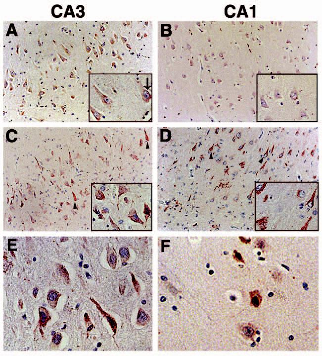
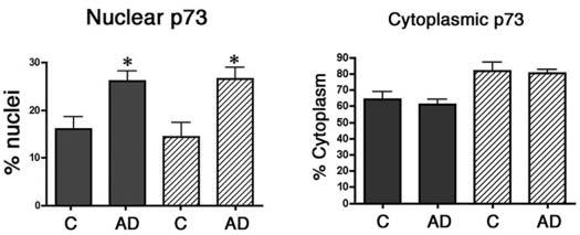
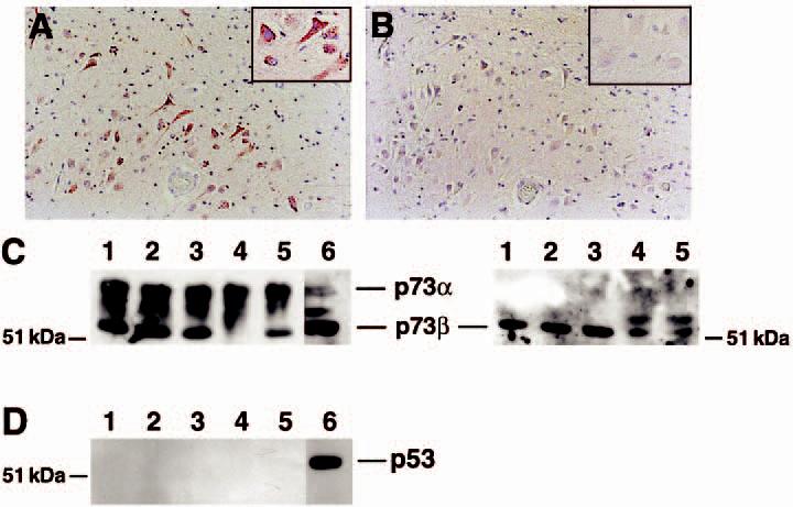
The molecular mechanisms that regulate neuronal survival vs. death during Alzheimer disease (AD) remain unclear. Nonetheless, a number of recent studies indicate that increased expression or altered subcellular distribution of numerous cell cycle proteins during AD may contribute to disease pathogenesis. Because homologues of p53, a key regulatory protein in the cell cycle, such as p73, have been identified and shown to participate in cellular differentiation and death pathways, we examined the expression and distribution of p73 in the hippocampus of eight control and 16 AD subjects. In control subjects, hippocampal pyramidal neurones exhibit p73 immunoreactivity that is distributed predominately in the cytoplasm. In AD hippocampus, increased levels of p73 are located in the nucleus of pyramidal neurones and p73 is located in dystrophic neurites and cytoskeletal pathology. Immunoblot analysis confirmed the presence of p73 in the hippocampus. These data indicate that p73 is expressed within hippocampal pyramidal neurones and exhibits altered subcellular distribution in AD.


Figures

Figure 1
Figure 1
p73 immunoreactivity in the CA3 and CA1 regions of the hippocampus using anti-p73 antibody H79. (A and B): p73 is located in neuronal cell bodies of the CA3 and CA1 hippocampal regions from control subjects. p73 is denoted in red and the sections counterstained with haematoxylin. Insets are high power magnification. (C and D): within the hippocampus of AD subjects, p73 is also detected in neurites and structures resembling neurofibrillary tangles (arrowheads). Each panel is ×200 magnification and the insets are ×400. (E and F): high power magnification (×400) of hippocampal pyramidal neurones immunolabelled for p73 in the CA3 (E) and CA1 (F) regions of AD brain. Panel (A) corresponds to case Control 8; panel (B) represents case Control 1; panel (C) is case AD 9; panel (D) is case AD 16; panel (E) is case AD 3; panel (F) is case AD 8 in Table 1. AD, Alzheimer disease.
Figure 2
Figure 2
The percentage of pyramidal neurones exhibiting p73 in the nucleus and cytoplasm of control (C) and Alzheimer disease (AD) brain. Pyramidal neurones within the CA1 and CA3 regions of the hippocampus were scored for the presence of p73 in the nucleus and cytoplasm as described in the section ‘Materials and methods’. Data represent the mean ± SEM for each group. Asterisks indicate significant difference in nuclear p73 between the control and AD groups (P < 0.008 for both CA1 and CA3).
Figure 3
Figure 3
p73 colocalizes to a subset of pyramidal neurones containing neurofibrillary tangles. (A–C): consecutive sections from Alzheimer disease (AD) hippocampus immunostained for Alz50 (A), p73 using the Ab-4 antibody (B), and beta-amyloid using 4G8 (C). Arrows indicate colocalization of p73 in tangle-bearing neurones and asterisks represent locations of amyloid-containing plaques. Magnification in panels A–C is ×200. (D and E): double-label confocal laser microscopy demonstrates colocalization of p73 (red) in tangle-bearing neurones (green) denoted using Alz50 antibody. Note that p73 as detected by the Ab-4 antibody is located in either the cytoplasm or nucleus (arrows) of these neurones. Bar corresponds to 20 μm in each panel. Panels (A–C) represent case AD 10, panel (D) represents case AD 13, and panel (E) represents case AD 6 in Table 1.
Figure 4
Figure 4
Abundant p73 protein levels in the hippocampus. Consecutive sections from the CA1 region of the hippocampus were immunolabelled for p73 using the Ab-4 antibody (A) or p53 (B). While p73 immunoreactivity was evident, little p53 protein was observed in the hippocampus from control or Alzheimer disease (AD) subjects. Panels (A) and (B) represent case AD 8 in Table 1. Magnification for both panels is ×200. Insets are high power magnification of the same region of the section. (C) Immunoblot for p73 (left) and p73β (right) using tissue extracts from the hippocampus of control and AD subjects. Lanes 1 and 2 represent control subjects and lanes 3–5 represent AD subjects (AD cases 12, 13, and 7, respectively). Lane 6 is a nuclear extract from cultured neuroblastoma cells as a positive control for p73. H79 antibody was used to detect p73 and C20 was used to detect p73β. Equal protein was loaded into each lane. (D) p53 protein was below detectable limits within the same tissue extracts. Identical lane assignments as in panel C.

Similar articles

Cited by

References

    1. Lyras L, Cairns N, Jenner A, Jenner P, Halliwell B. An assessment of oxidative damage to proteins, lipids, and DNA in brain from patients with Alzheimer's disease. J Neurochem. 1997;63:2061–9. - PubMed
    1. Smith MA, Rottkamp CA, Nunomura A, Raina AK, Perry G. Oxidative stress in Alzheimer's disease. Biochim Biophys Acta. 2000;1502:139–44. - PubMed
    1. Smith MA, Sayre LM, Anderson VE, Beal MF, Kowall N, Richey PL, Perry G. Oxidative damage in Alzheimer's disease. Nature. 1996;382:120–1. - PubMed
    1. Bennett M, Macdonald K, Chan S-W, Luzio JP, Simari R, Weissberg P. Cell surface trafficking of Fas. A rapid mechanism of p53-mediated apoptosis. Science. 1998;282:290–3. - PubMed
    1. De Laurenzi V, Melino G. Evolution of functions within the p53/p63/p73 family. Ann NY Acad Sci. 2000;926:90–100. - PubMed

Publication types

MeSH terms