

Review
doi: 10.1016/j.molmed.2003.11.007.
TGF-beta, T-cell tolerance and anti-CD3 therapy
Affiliations
- PMID: 14720578
- PMCID: PMC2796480
- DOI: 10.1016/j.molmed.2003.11.007
Item in Clipboard
Review
TGF-beta, T-cell tolerance and anti-CD3 therapy
Trends Mol Med.
2004 Jan.
Abstract
Recent studies by Chatenoud and co-workers suggest that non-mitogenic F(ab′)2 fragments of anti-CD3 antibodies, which cannot bind the Fc receptor, induce a prolonged period of tolerance and prevent diabetes in nonobese diabetic (NOD) mice. Tolerance is established by regulatory T cells through the production of transforming growth factor-β1 (TGF-β1), but the mechanism by which TGF-β1 confers this activity is unclear. Analysis of mice deficient in TGF-β1 suggests that TGF-β1 raises the threshold at which intracellular calcium activates T cells to a level that prevents an autoimmune response.
Figures
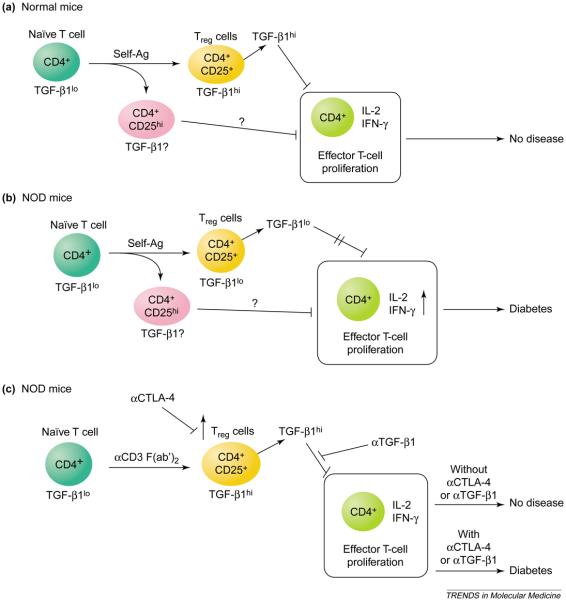
Regulatory T (Treg) cells prevent diabetes in nonobese diabetic (NOD) mice. (a) CD4+CD25+ T cells develop in the thymus and prevent peripheral T-cell activation and autoimmune disease. Treg cells inhibit proliferation and cytokine production by T helper (Th) cells on stimulation in vitro and in vivo. On stimulation, Treg cells produce transforming growth factor-β1 (TGF-β1), which in turn inhibits the activation of Th cells. CD4+CD25hi Treg cells also inhibit Th-cell responses in a TGF-β1-independent manner, although their role in vivo is still unclear. (b) Deficiency in functional CD4+CD25+ Treg cells causes diabetes in NOD mice. In these mice, Th cells are spontaneously activated and Treg cells do not produce TGF-β1, thereby resulting in autoimmune type 1 diabetes. (c) Induction of functional Treg cells prevents diabetes in NOD mice through TGF-β1-mediated inhibition of Th-cell activation. Treatment of NOD mice with anti-CD3 generates functional CD4+CD25+ Treg cells from CD4+ CD25− T cells. Blockade of cytotoxic T-lymphocyte-associated antigen 4 (CTLA-4)-mediated co-stimulation inhibits the production of TGF-β1 by Treg cells and thus reverses the protection mediated by anti-CD3 treatment. Consequently, blockade by anti-TGF-β1 also reverses the protection mediated by Treg cells.
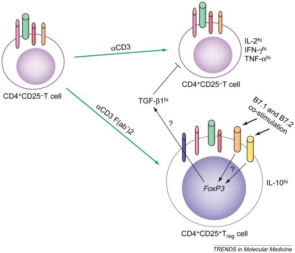
Production of transforming growth factor-β1 (TGF-β1) is essential for the function of regulatory T (Treg) cells. On activation of CD4+CD25− T cells by anti-CD3, T helper (Th) cells produce pro-inflammatory cytokines such as interleukin (IL)-2, interferon-γ (IFN-γ) and tumor necrosis factor-α (TNF-α). The anti-inflammatory cytokines TGF-β1 and IL-10 (not shown) are also induced to a lesser extent. Treatment of CD4+CD25− T cells with the non-mitogenic anti-CD3 F(ab′)2 fragment induces expression of CD25, resulting in CD4+CD25+ Treg cells. Activated Treg cells produce mainly TGF-β1 and IL-10, but do not produce IL-2, IFN-γ or TNF-α. Co-stimulation through CD28 is essential for the development of Treg cells and their survival, whereas co-stimulation through cytotoxic T-lymphocyte-associated antigen 4 (CTLA-4) is essential for their functional response. Expression of the transcription factor gene Foxp3 is essential for the development and function of Treg cells, which inhibit Th-cell activation through the juxtacrine activity of TGF-β1. Blue cylinder, CD25; orange cylinder, CD28; pink cylinder, CD4; yellow cylinder, CTLA-4; green cylinder, T-cell receptor; red cylinder, CD3.
References
-
- Belghith M, et al. TGF-β-dependent mechanisms mediate restoration of self-tolerance induced by antibodies to CD3 in overt autoimmune diabetes. Nat. Med. 2003;9:1202–1208. - PubMed
-
- Stockinger B. T lymphocyte tolerance: from thymic deletion to peripheral control mechanisms. Adv. Immunol. 1999;71:229–265. - PubMed
-
- Bonomo A, et al. Premature escape of double-positive thymocytes to the periphery of young mice. Possible role in autoimmunity. J. Immunol. 1994;152:1509–1514. - PubMed
-
- Wood KJ, Sakaguchi S. Regulatory T cells in transplantation tolerance. Nat. Rev. Immunol. 2003;3:199–210. - PubMed
-
- Shevach EM. CD4+CD25+ suppressor T cells: more questions than answers. Nat. Rev. Immunol. 2002;2:389–400. - PubMed
Publication types
MeSH terms
Substances
Grants and funding
LinkOut - more resources
Full Text Sources
Medical
Miscellaneous
