. 2004 Feb;78(3):1069-79.
doi: 10.1128/jvi.78.3.1069-1079.2004.

The membrane-proximal tyrosine-based sorting signal of human immunodeficiency virus type 1 gp41 is required for optimal viral infectivity

Affiliations

The membrane-proximal tyrosine-based sorting signal of human immunodeficiency virus type 1 gp41 is required for optimal viral infectivity

John R Day et al. J Virol. 2004 Feb.

Abstract

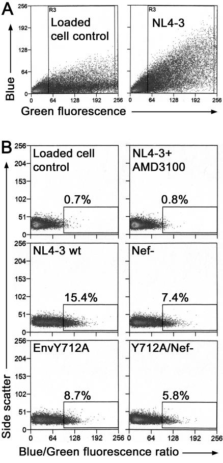
The membrane-proximal tyrosine-based sorting motif in the cytoplasmic domain of the human immunodeficiency virus type 1 Env glycoprotein is important for endocytosis from the plasma membrane, basolateral targeting of viral budding in polarized epithelial cells, and polarized budding from a localized region of the lymphocyte plasma membrane. To study the role of the Env sorting motif (Y712XXL) in infectivity, the incorporation of Env into virions, and viral entry, we disrupted the motif with a tyrosine-to-alanine substitution. To investigate the relationship between the Env sorting motif and the enhancement of infectivity by Nef, the EnvY712A substitution was made in both Nef-positive and Nef-negative backgrounds. In spreading infections, including those using primary lymphocytes, the growth of the Y712A mutant was as impaired as Nef-negative virus, and the EnvY712A/Delta-Nef combination mutant was almost completely defective. In single-round infections using CD4-positive HeLa cells, the EnvY712A mutation impaired infectivity, and Nef retained the ability to enhance the infectivity in the context of EnvY712A. EnvY712 and Nef were required for the optimal infectivity of virions produced from either HEK293T or MT4 cells, but these sequences were required for the optimal incorporation of Env only when virions were produced from MT4 cells. Despite the wild-type levels of Env in viruses produced from 293T cells, the entry of the EnvY712A and Delta-Nef mutants into target cells was impaired. We conclude that the membrane-proximal tyrosine-based sorting motif of gp41 Env is, like Nef, important for optimal viral infectivity and, in the case of MT4 T cells, virion incorporation of Env. Nef does not require the Y712XXL motif to enhance viral infectivity. The finding that EnvY712 and Nef each affect the efficiency of viral entry independently of the Env content of virions suggests that both viral proteins are involved in trafficking events that influence morphogenesis to produce maximally fusogenic virus.


Figures

FIG. 1.
FIG. 1.
Schematic representation of the HIV-1 envelope precursor glycoprotein (gp160). The surface subunit, gp120, and transmembrane subunit, gp41, are indicated. The gp41 subunit consists of an extracellular domain, transmembrane domain (TM), and a cytoplasmic domain. Membrane-proximal amino acids of the cytoplasmic domain of HIV-1 clone NL4-3 are shown beneath the schematic, with the membrane-proximal tyrosine-based sorting motif underlined. The tyrosine at residue 712 (in boldface type) was replaced with alanine by PCR site-directed mutagenesis.
FIG. 2.
FIG. 2.
Expression of Env in MT4 and HeLa cells. (A) Western blot of MT4 cell lysates for gp160 and p24. MT4 cells were transfected with wild-type and mutant proviruses and pelleted 3 days later. Cells were washed in PBS and then resuspended in reducing SDS-PAGE sample buffer. Equal amounts of p24 capsid per lane were run on an SDS-polyacrylamide gel, transferred to a PVDF membrane, probed sequentially with sheep antiserum to HIV-1 gp120 (DV-012) and mouse anti-p24 (7A8.1), and developed with a chemiluminescent substrate. Bands corresponding to gp160 and p24 are indicated. (B) Immunofluorescence of HeLa-CD4 cells. Cells were transfected with wild-type NL4-3 or EnvY712A provirus, fixed 2 days later, permeabilized, and stained with mouse anti-gp41 followed by a Rhodamine-X-conjugated secondary antibody. Slides were analyzed with a Zeiss Axioskop fluorescent microscope (magnification, ×400), and images were processed using Adobe Photoshop.
FIG. 3.
FIG. 3.
Growth of viruses in PBMC and CEM and MT4 cells. Viruses were produced by transfection of HEK293 cells. One million PBMC or CEM or MT4 cells were infected in duplicate with viruses normalized by p24 capsid protein concentration. Samples of the cultures were taken over time and analyzed for p24 protein concentration by ELISA. CEM and MT4 cells were split 1:4 after every culture sampling. Each experiment was performed at least twice, each in duplicate, with similar results in both the hierarchy and extent of replication. (A) Primary PBMCs were stimulated with phytohemagglutinin for 3 days prior to infection with virus containing 50 ng of p24 capsid, supplemented with IL-2. (B) CEM T cells were infected with 250 ng of p24 capsid. (C) MT4 T cells were infected with 5 ng of p24 capsid. Error bars indicate standard deviations of duplicate infections.
FIG. 4.
FIG. 4.
Infectivity of HEK293T- and MT4-produced virus. (A) Viruses were produced by transfection of either 293T or MT4 cells as indicated underneath the x axis. Virus-containing supernatants normalized by p24 capsid were added to P4.R5 indicator cells that contain β-galactosidase under control of the HIV-1 LTR. Following 2 days of incubation, cells were stained with X-Gal and blue foci were counted. Infectivity data are expressed as a ratio of the number of blue cells per ng of p24 inoculum normalized to wild type. The 293T data shown are the averages of duplicates, and the MT4 data shown are the averages between two independent assays. (B) Infectivity of 293-produced virus compared in two distinct assays as indicated underneath the x axis. The P4 assay was performed as described in panel A. For the syncytium induction assay, HeLa-CD4 cells (clone 1022) were infected with viruses normalized by p24 capsid concentration and incubated for 3 days. Cells were stained with crystal violet, and the syncytia were counted. Data shown are the averages of duplicates. (C) Infectivity of various 293T-produced EnvY712 mutants tested in the P4 infectivity assay as above. Data shown are the averages of duplicates. Error bars indicate standard deviation.
FIG. 5.
FIG. 5.
Env incorporation in MT4-produced virus. (A) Plot of band density versus nanograms of recombinant gp120. Known amounts of recombinant gp120 were loaded onto an SDS-polyacrylamide gel, transferred to a PVDF membrane, probed with sheep antiserum to HIV-1 gp120, and developed with a chemiluminescent substrate. Bands were analyzed using ImageJ (version 1.29t) software (Wayne Rasband, National Institutes of Health). (Inset: Western blot image of recombinant gp120.) (B) Western blot of MT4-produced virus. Virus-containing supernatants produced from transfected MT4 cells were filtered through 0.2-μm-pore-size filters and centrifuged at 23,500 × g for 2 h at 4°C. Viral pellets were then resuspended in a small volume of reducing SDS-PAGE sample buffer. Viral pellet suspensions were normalized for p24 concentration and electrophoresed and electroblotted as described in Fig. 2A. Bands corresponding to gp120 and p24 are indicated. (C) Relative amounts of gp120 incorporation. gp120 and p24 bands were scanned and analyzed using ImageJ (version 1.29t) software. Band densities are expressed as a ratio of gp120 to p24 and are an average of two independent experiments. Error bars indicate standard deviation.
FIG. 6.
FIG. 6.
Envelope incorporation in HEK293T-produced virus. Virus-containing supernatants produced from transfected HEK293T cells were pelleted and analyzed as described for Fig. 5B. Bands corresponding to gp160, gp120, and p24 are indicated.
FIG. 7.
FIG. 7.
β-Lactamase-Vpr entry assay. Viruses were produced by cotransfection of the indicated provirus and pMM310, a β-lactamase-Vpr (BlaM-Vpr) fusion protein expression vector. Viruses were pelleted, resuspended in 1 ml of medium, normalized by p24 capsid concentration, and then used to infect P4.R5 cells for 5 h. After washing, cells were incubated with the β-lactamase-sensitive fluorochrome CCF2/AM for 16 to 18 h at 25°C. Analysis was performed by flow cytometry. Uncleaved substrate was detected as green fluorescence, and cleaved substrate was detected as blue fluorescence. (A) Plot of green fluorescence versus blue fluorescence. Loaded cell control refers to uninfected cells loaded with CCF2/AM. Box R3 encloses cells used for the ratio analysis in panel B, cells positive for CCF2/AM loading. (B) Data are expressed as a ratio of blue to green fluorescence versus side scatter and are gated using uninfected cells (loaded cell control). AMD3100 (100 nm) is a CXCR4 antagonist used to block entry into cells.
FIG. 8.
FIG. 8.
Entry assay summary and β-lactamase activity. (A) Data from three independent experiments as shown in Fig. 7 were expressed as a percentage of wild-type entry and averaged. (B) β-Lactamase activity of BlaM-Vpr-containing viruses. Pelleted viruses normalized by p24 capsid concentration were incubated with the chromogenic substrate nitrocefin for 22 h at room temperature. Samples were analyzed in a microplate spectrophotometer at 490 nm. Nitrocefin diluted into sample diluent was measured as background, and 20 μl of an ampicillin-resistant bacterial culture was used as a positive control. Error bars, standard deviations.

References

    1. Aiken, C., and D. Trono. 1995. Nef stimulates human immunodeficiency virus type 1 proviral DNA synthesis. J. Virol. 69:5048-5056. - PMC - PubMed
    1. Akari, H., T. Fukumori, and A. Adachi. 2000. Cell-dependent requirement of human immunodeficiency virus type 1 gp41 cytoplasmic tail for Env incorporation into virions. J. Virol. 74:4891-4893. - PMC - PubMed
    1. Berlioz-Torrent, C., B. L. Shacklett, L. Erdtmann, L. Delamarre, I. Bouchaert, P. Sonigo, M. C. Dokhelar, and R. Benarous. 1999. Interactions of the cytoplasmic domains of human and simian retroviral transmembrane proteins with components of the clathrin adaptor complexes modulate intracellular and cell surface expression of envelope glycoproteins. J. Virol. 73:1350-1361. - PMC - PubMed
    1. Blot, G., K. Janvier, S. Le Panse, R. Benarous, and C. Berlioz-Torrent. 2003. Targeting of the human immunodeficiency virus type 1 envelope to the trans-Golgi network through binding to TIP47 is required for Env incorporation into virions and infectivity. J. Virol. 77:6931-6945. - PMC - PubMed
    1. Boge, M., S. Wyss, J. S. Bonifacino, and M. Thali. 1998. A membrane-proximal tyrosine-based signal mediates internalization of the HIV-1 envelope glycoprotein via interaction with the AP-2 clathrin adaptor. J. Biol. Chem. 273:15773-15778. - PubMed

Publication types

MeSH terms