. 2004 Feb;78(3):1093-100.
doi: 10.1128/jvi.78.3.1093-1100.2004.

Novel insect picorna-like virus identified in the brains of aggressive worker honeybees

Affiliations

Novel insect picorna-like virus identified in the brains of aggressive worker honeybees

Tomoko Fujiyuki et al. J Virol. 2004 Feb.

Abstract

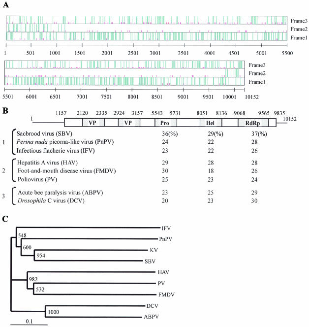
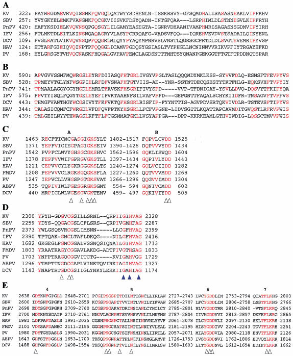
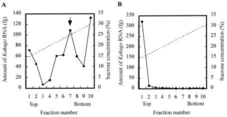
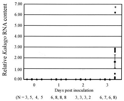
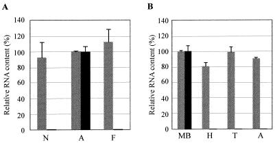
To identify candidate genes involved in the aggressive behavior of worker honeybees, we used the differential display method to search for RNAs exclusively detected in the brains of aggressive workers that had attacked a hornet. We identified a novel, 10,152-nucleotide RNA, termed Kakugo RNA. Kakugo RNA encodes a protein of 2,893 amino acid residues that shares structural features and sequence similarities with various picorna-like virus polyproteins, especially those from sacbrood virus, which infects honeybees. The Kakugo protein contains several domains that correspond to the virion protein, helicase, protease, and RNA-dependent RNA polymerase domains of various picorna-like virus polyproteins. When the worker bee tissue lysate was subjected to sucrose density gradient centrifugation, Kakugo RNA, except for the material at the bottom, was separated into two major peaks. One of the peaks corresponded to the position of Kakugo mRNA, and the other corresponded to the position of the poliovirus virion. These results suggest that the Kakugo RNA exists as an mRNA-like free RNA and virion RNA in the honeybee. Furthermore, injection of the lysate supernatant from the attacker heads into the heads of noninfected bees resulted in a marked increase in Kakugo RNA. These results demonstrate that Kakugo RNA is a plus-strand RNA of a novel picorna-like virus and that the brains of aggressive workers are infected by this novel virus. Kakugo RNA was detected in aggressive workers but not in nurse bees or foragers. In aggressive workers, Kakugo RNA was detected in the brain but not in the thorax or abdomen, indicating a close relation between viral infection in the brain and aggressive worker behaviors.


Figures

FIG. 1.
FIG. 1.
Identification of Kakugo RNA in the brains of attacker honeybees. (A) A hornet (blue arrow) was hung by a thread and presented as a decoy to the guard bees. (B) Some guard bees scrambled and obstinately attacked the hornet. (C) The hornet was presented to the workers inside the hive. (D) Some of the workers escaped from it. (E) Semiquantitative RT-PCR using total RNA from the MBs of the escapers (lane 1) and attackers (lane 2). Gene-specific primer sets for Kakugo and actin were used. RT (−), control experiment with RT-negative template and Kakugo-specific primer set.
FIG. 2.
FIG. 2.
Kakugo RNA encodes a polyprotein of a putative picorna-like virus. (A) Graphic representation of the positions of the initiation codons and termination codons in the Kakugo cDNA. Termination codons (TAA, TAG, and TGA) and the initiation codon (ATG) are indicated by long blue and short pink vertical lines, respectively, in each of three reading frames. The numbers indicate base numbers from the 5′ end. (B) Structure of Kakugo cDNA and comparison of putative amino acid sequence of the Kakugo protein with those of other picorna-like virus polyproteins. Open and shaded boxes show the ORFs carried by the Kakugo cDNA and the domains of the Kakugo polyprotein, respectively. Numbers indicate the base positions corresponding to each domain. Sequence identities with the Kakugo protein in the helicase (Hel), protease (Pro), and RdRp domains are indicated below the corresponding domains. Bracket 1, the insect picorna-like viruses SBV (8), Perina nuda picorna-like virus (PnPV) (37), and infectious flacherie virus (IFV) (13); bracket 2, the mammalian picornaviruses hepatitis A virus (HAV) (22), foot-and-mouth disease virus (FMDV) (7), and PV (23); bracket 3, the cricket paralysis-like viruses acute bee paralysis virus (ABPV) (10) and Drosophila C virus (DCV) (16). (C) Phylogenetic tree constructed with the highly conserved amino acid sequences encompassing motifs 1 to 8 of the RdRp domains by the neighbor-joining method. KV, putative Kakugo virus. Numbers at each node represent bootstrap values as the results of 1,000 replicons.
FIG. 3.
FIG. 3.
Southern blotting analysis of Kakugo DNA. Southern blotting analysis was performed with honeybee genomic DNA (10 μg) digested with EcoRI, with a partial Kakugo cDNA as a probe (lane 1). The Mblk-1 gene, which encodes a transcription factor (26, 27, 32), was also analyzed as a loading control (lane 2). Plasmid cDNAs (5 pg each) for Kakugo and Mblk-1 were included as hybridization controls (bottom). The band positions are indicated by arrows.
FIG. 4.
FIG. 4.
Comparison of the amino acid sequences of functional domains of the Kakugo polyprotein and other picorna-like virus polyproteins. (A) One of the VP regions of Kakugo virus with the corresponding region of SBV, CP1 of PnPV, VP3 of IFV, VP1 of DCV, VP2 of HAV, and the corresponding region of PV. Residues that are identical for at least four of the seven viruses are shown in red (A and B). (B) The other VP region of the Kakugo virus with the corresponding region of SBV, CP3 of PnPV, VP1 of IFV, VP2 of DCV, VP3 of HAV, and the corresponding region of PV. (C) The putative motifs A and B in the helicase domain of the Kakugo virus with those of SBV, PnPV, IFV, HAV, FMDV, PV, ABPV, and DCV. Amino acid residues that are essential for enzymatic activity are indicated by arrowheads (C and E). (D) The C terminus of the putative protease domain of Kakugo virus with those of SBV, PnPV, IFV, HAV, FMDV, PV, ABPV, and DCV. The protease motif and amino acid residues involved in substrate binding are indicated with white and blue arrowheads, respectively. (E) The putative motifs 4 through 7 in the RdRp domains of Kakugo virus with those of SBV, PnPV, IFV, HAV, FMDV, PV, ABPV, and DCV. Residues that are identical in at least five of the nine viruses are shown in red (C, D, and E). Arrowheads indicate the consensus amino acids within RdRp motifs. Numbers show amino acid positions from the N terminus.
FIG. 5.
FIG. 5.
Sucrose density gradient centrifugation of the honeybee tissue lysate that contained Kakugo RNA. The lysate (A) and the total RNA (B) prepared from thoraxes of the foragers inoculated with Kakugo virus were each subjected to sucrose density gradient centrifugation, and the amount of the Kakugo RNA in each fraction was determined by quantitative RT-PCR. Arrow, the fraction in which the PV virion was mainly found. The solid and broken lines indicate the amount of Kakugo RNA and the putative sucrose concentration, respectively.
FIG. 6.
FIG. 6.
Kakugo RNA constitutes an infectious virus. Foragers inoculated either with head lysate from the attackers (circle), head lysate from the foragers (triangle), or PBS (square) were collected at different times after inoculation. Noninjected foragers (lozenge) were also collected as controls. Kakugo RNA and actin mRNA levels, which served as a control, were determined by quantitative RT-PCR. The mean value of relative Kakugo RNA content normalized to that of actin mRNA is shown with a bold bar showing standard errors. N, the number of samples used for each experiment; asterisk, differences were significant, with a P value of <0.05 by the unpaired t test.
FIG. 7.
FIG. 7.
Kakugo RNA is detected almost exclusively in the brains of attackers. (A) Relative Kakugo RNA content in the MBs of nurse bees (N), attackers (A), and foragers (F). As many as 100 of each were examined by quantitative RT-PCR with Kakugo-specific primers and probes (black bar). actin mRNA was also examined to show that essentially the same amount of RNA was contained in each sample (dotted bars). Values are represented as percentages of Kakugo or actin mRNA relative to that in the attacker brains. (B) Relative RNA content of the MBs, heads (H), thoraxes (T), and abdomens (A) from five attackers, examined with Kakugo-specific (black bar) and actin-specific (dotted bars) primers and probes.

References

    1. Bailey, L. 1969. The multiplication and spread of sacbrood virus of bees. Ann. Appl. Biol. 63:483-491. - PubMed
    1. Bailey, L., and E. F. W. Fernando. 1972. Effects of sacbrood virus on adult honey-bees. Ann. Appl. Biol. 72:27-35.
    1. Bowen-Walker, P. L., S. J. Martin, and A. Gunn. 1999. The transmission of deformed wing virus between honeybees (Apis mellifera L.) by the ectoparasitic mite Varroa jacobsoni Oud. J. Invertebr. Pathol. 73:101-106. - PubMed
    1. Breed, M. D., G. E. Robinson, and J. R. E. Page. 1990. Division of labor during honey bee colony defense. Behav. Ecol. Sociobiol. 27:395-401.
    1. Breed, M. D., T. A. Smith, and A. Torres. 1992. Role of guard honey bees (Hymenoptera: Apidae) in nestmate discrimination and replacement of removed guards. Ann. Entomol. Soc. 85:633-637.

Publication types

MeSH terms

Associated data

LinkOut - more resources