. 2004 Feb;78(3):1109-20.
doi: 10.1128/jvi.78.3.1109-1120.2004.

Downregulation of major histocompatibility complex class I by human ubiquitin ligases related to viral immune evasion proteins

Affiliations

Downregulation of major histocompatibility complex class I by human ubiquitin ligases related to viral immune evasion proteins

Eric Bartee et al. J Virol. 2004 Feb.

Abstract

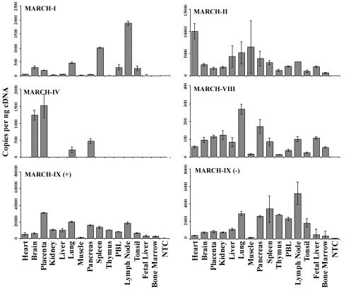
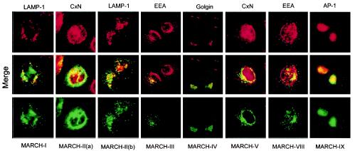
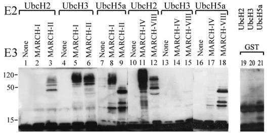
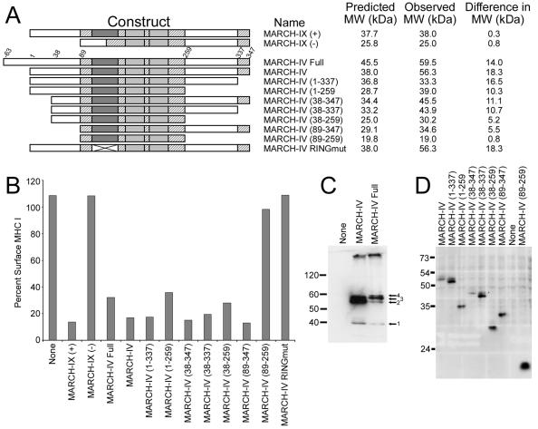
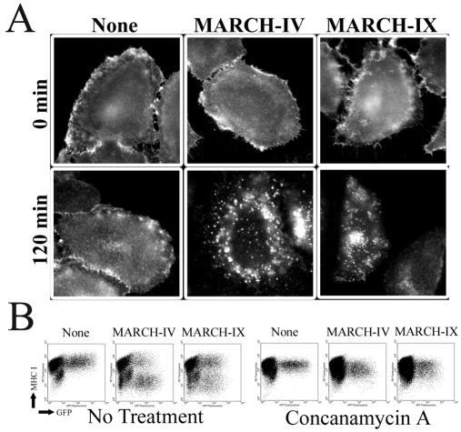
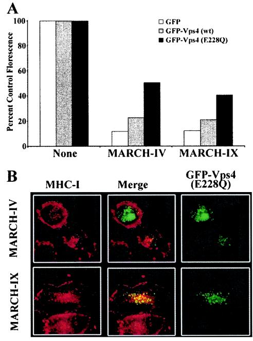
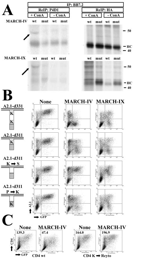
Poxviruses and gamma-2 herpesviruses share the K3 family of viral immune evasion proteins that inhibit the surface expression of glycoproteins such as major histocompatibility complex class I (MHC-I), B7.2, ICAM-1, and CD95(Fas). K3 family proteins contain an amino-terminal PHD/LAP or RING-CH domain followed by two transmembrane domains. To examine whether human homologues are functionally related to the viral immunoevasins, we studied seven membrane-associated RING-CH (MARCH) proteins. All MARCH proteins located to subcellular membranes, and several MARCH proteins reduced surface levels of known substrates of the viral K3 family. Two closely related proteins, MARCH-IV and MARCH-IX, reduced surface expression of MHC-I molecules. In the presence of MARCH-IV or MARCH-IX, MHC-I was ubiquitinated and rapidly internalized by endocytosis, whereas MHC-I molecules lacking lysines in their cytoplasmic tail were resistant to downregulation. The amino-terminal regions containing the RING-CH domain of several MARCH proteins examined catalyzed multiubiquitin formation in vitro, suggesting that MARCH proteins are ubiquitin ligases. The functional similarity of the MARCH family and the K3 family suggests that the viral immune evasion proteins were derived from MARCH proteins, a novel family of transmembrane ubiquitin ligases that seems to target glycoproteins for lysosomal destruction via ubiquitination of the cytoplasmic tail.


Figures

FIG. 1.
FIG. 1.
Human homologues of viral K3 family proteins. The RING-CH domain from the nine identified human MARCH proteins (I to IX) and the known viral K3 family proteins were aligned by using Vector NTI 7.0 software. The RING-CH domain is shown boxed in dark gray, whereas other conserved amino acids are shown boxed in light gray. The hydropathy plots (middle panel) for each MARCH protein were generated by using TMPRED (www.ch.embnet.org). The location of the RING-CH domain is shown by a black box under the hydropathy plot, whereas the number of transmembrane domains predicted is shown to the right of the RING-CH alignment. Phylogenetic relationships for the full-length sequences (bottom) were generated by using Vector NTI suite 7 software.
FIG. 2.
FIG. 2.
Tissue distribution of MARCH proteins. Expression of MARCH-I, -II, -IV, -VIII, and -IX was measured by real-time PCR in a panel of human tissue cDNAs (Clontech). MARCH-IX occurs in two splice variants, one of them missing the RING-CH domain. Both variants were included in this analysis. Expression of each gene was quantified by using absolute standards created from plasmid DNA. Concentrations of cDNAs were normalized by using the housekeeping genes GAPDH (glyceraldehyde-3-phosphate dehydrogenase) and β-actin.
FIG. 3.
FIG. 3.
Localization of MARCH proteins to intracellular membranous organelles. HeLa cells were transfected with plasmids encoding C-terminally Flag-tagged versions of the indicated MARCH proteins. At 20 h posttransfection, cells were stained with antibodies against indicated cellular markers (top panel in red) or anti-FLAG (bottom panel in green). Colocalization is visualized as yellow in the merged panel (middle). All cellular markers were examined against all MARCH proteins. However, only results that indicated a significant overlap are shown. The markers of subcellular compartments were calnexin (Cxn) for ER, Golgin-97 for Golgi, Adaptor protein 1 (AP-1) for the trans-Golgi network, early endosomal antigen (EEA) for endosomes, and LAMP-1 for lysosomes.
FIG. 4.
FIG. 4.
MARCH proteins act as ubiquitin ligases in vitro. GST fusion proteins of the amino-terminal domains of individual MARCH proteins were incubated with ubiquitin, ATP, E1, and the indicated ubiquitin-conjugating enzymes (ubc, E2) and separated by SDS-polyacrylamide gel electrophoresis prior to immunoblotting with the ubiquitin-specific antibody P4D1. Ubiquitin ligase activity is indicated by the appearance of high-molecular-weight ubiquitinated species. Ubiquitinated proteins were not detected in the absence of E3s (None) or with GST alone (unpublished data).
FIG. 5.
FIG. 5.
Downregulation of surface glycoproteins by the MARCH family. (A) HeLa cells were transfected with the respective MARCH expression plasmid and GFP. Surface expression of indicated surface proteins was measured via flow cytometry with specific primary antibodies and a phycoerythrin-conjugated secondary at 24 h posttransfection (unpublished data). Black arrows indicate significant downregulation. Expression of FLAG-tagged versions of MARCH proteins was confirmed with anti-FLAG antibody by immunoprecipitation (B), whereas steady-state levels were measured by immunoblot (C). Introduction of the FLAG tag at the carboxy terminus of MARCH proteins did not affect the ability of these proteins to downregulate their substrates (data not shown).
FIG. 6.
FIG. 6.
Functional domains of MARCH-IV and MARCH-IX. (A) Diagram showing the homology between MARCH-IV and MARCH-IX, as well as the deletion mutants in MARCH-IV. The RING-CH domain (dark gray), the two transmembrane domains (light gray) and additional regions (hatched) are > 90% identical, whereas the remaining parts of the molecules (white) are <20% conserved. MARCH-IX (−) represents a splice variant lacking the RING-CH domain. Two conserved cysteines in the RING-CH domain were replaced with serines in the MARCH-IV RING mutant. N- and C-terminal deletion constructs of MARCH-IV are indicated; the predicted full-length construct contains a 63-amino-acid N-terminal extension. (B) Ratio (in percent) of the mean fluorescence of surface MHC-I detected by flow cytometry with W6/32 after 24 h of MARCH-transfected versus nontransfected HeLa cells gated for GFP. Both RING-CH mutants were unable to downregulate MHC surface expression. All MARCH-IV deletion constructs reduced MHC-I surface expression except MARCH-IV (89-259) containing the RING-CH and transmembrane regions only. (C) Immunoprecipitation, followed by immunoblotting with anti-FLAG, reveals the presence of two major protein bands of ca. 55 kDa (arrows 2 and 3) in cells transfected with both full-length MARCH-IV and MARCH-IV (1-347), whereas the predicted full-length protein (arrow 4) is only a minor species. (D) Immunoblot with anti-FLAG reveals the observed MW of the truncated MARCH-IV proteins. The difference between observed and predicted molecular weight is shown in panel A. Only MARCH-IV (89-259) migrated at the predicted position.
FIG. 7.
FIG. 7.
Endocytosis and lysosomal degradation of MHC-I by MARCH-IV and MARCH-IX. (A) Uptake of MHC-I in HeLa cells transfected with vector control, MARCH-IV or MARCH-IX. At 24 h posttransfection, MHC-I was stained with antiserum K455 and either fixed after 30 min at 4°C (top panel) or transferred to 37°C for 2 h (bottom panel) prior to fixation and staining with Alexafluor 594 anti-rabbit secondary antibody. (B) Flow cytometry of MHC-I (W6/32 staining) on MARCH-IV or IX-transfected HeLa cells in the presence or absence of concanamycin A.
FIG. 8.
FIG. 8.
Vps4 restores MHC-I surface levels and colocalizes with MHC-I in HeLa cells transfected with MARCH-IV and MARCH-IX. (A) HeLa cells were transfected with GFP only or GFP-tagged wild-type Vps4 or the dominant-negative, ATP-hydolysis-deficient mutant GFP-Vps4(E228Q) (5), together with vector control MARCH-IV or MARCH-IX. At 24 h after transfection, cells were stained with W6/32, and the mean fluorescence of GFP-positive cells was determined. The ratio of the mean fluorescence of MARCH-transfected versus the corresponding vector-transfected cells is shown as a percentage. Cotransfection of GFP-Vps4(E228Q) partially restored MHC-I surface levels in MARCH-IV- and MARCH-IX-transfected cells compared to cells that were only transfected with GFP-Vps4(E228Q) and vector. (B) Uptake and colocalization of MHC-I molecules with GFP-Vps4(E228Q). Internalization of MHC-I by MARCH-IV and -IX was monitored as for Fig. 6, except that cells were cotransfected with GFP-Vps4(E228Q). Punctate staining of Vps4 (right panel, green) is consistent with its localization to endosomes (5). MHC-I (K455 staining, red, right panel) remained mostly at the cell surface in cells expressing high levels of GFP-Vps4(E228Q) (upper left cell in upper panel), whereas MHC-I and Vps4 colocalized in cells with lower GFP fluorescence, indicating lower Vps4 levels (cell in the center of upper and lower panel; merged fluorescence is shown in the middle column). No colocalization of MHC-I and Vps4 was observed in the absence of MARCH-IV and -IX, and wild-type Vps4 stained the cytoplasm (data not shown).
FIG. 9.
FIG. 9.
Ubiquitination of MHC-I and role of C-terminal lysines for MHC-I downregulation by MARCH-IV and MARCH-IX. (A) HeLa cells were transiently transfected with HA-tagged HLA-A2.1 d331 (31) and MARCH-IV, MARCH-IX, or their respective RING-CH mutants. MARCH-IX expression was fully induced by removal of tetracycline, whereas MARCH-IV expression was partially repressed with 5 ng of tetracycline since high levels of expression were not compatible with the long labeling protocol. Therefore, there is only a slight reduction of MHC expression levels in MARCH-IV transfectants compared to MARCH-IX transfectants. Where indicated, cells were treated with concanamycin A prior to and during labeling for 6 h. HLA-A2.1 was immunoprecipitated with BB7.2 and, after denaturation with SDS, reimmunoprecipitated with ubiquitin-specific antibody P4D1 (90% of the lysate, left panel) or anti-HA antibody (10% of the lysate, right panel). Due to the large amount of MHC heavy chain in the primary immunoprecipitation, some of it was carried over in the P4D1 reimmunoprecipitation. The arrow indicates ubiquitinated heavy chain. (B) Downregulation of HLA-A2.1 lysine mutants by MARCH-IV and MARCH-IX. Cytoplasmic tails of HLA-A2.1 are schematically depicted on the left (28). The number of lysines in the A2.1 tail (white) or the HA tag (gray) is shown. Surface expression of the individual constructs was monitored by flow cytometry with antibody BB7.2. Transfection with vector (left), MARCH-IV (middle) or MARCH-IV was tracked by cotransfection of GFP. (C) Surface expression of wild-type CD4 or CD4 lacking lysines in its cytoplasmic tail in the presence of MARCH-IV. Mean fluorescence values for GFP-positive cells are shown in the upper left.

References

    1. Aasland, R., T. J. Gibson, and A. F. Stewart. 1995. The PHD finger: implications for chromatin-mediated transcriptional regulation. Trends Biochem. Sci. 20:56-59. - PubMed
    1. Aravind, L., L. M. Iyer, and E. V. Koonin. 2003. Scores of RINGS but no PHDs in ubiquitin signaling. Cell Cycle 2:123-126. - PubMed
    1. Babst, M., D. J. Katzmann, E. J. Estepa-Sabal, T. Meerloo, and S. D. Emr. 2002. Escrt-III: an endosome-associated heterooligomeric protein complex required for mvb sorting. Dev. Cell 3:271-282. - PubMed
    1. Biederer, T., C. Volkwein, and T. Sommer. 1997. Role of Cue1p in ubiquitination and degradation at the ER surface. Science 278:1806-1809. - PubMed
    1. Bishop, N., and P. Woodman. 2000. ATPase-defective mammalian VPS4 localizes to aberrant endosomes and impairs cholesterol trafficking. Mol. Biol. Cell 11:227-239. - PMC - PubMed

Publication types