. 2004 Feb;78(3):1121-8.
doi: 10.1128/jvi.78.3.1121-1128.2004.

Nuclear localization but not PML protein is required for incorporation of the papillomavirus minor capsid protein L2 into virus-like particles

Affiliations

Nuclear localization but not PML protein is required for incorporation of the papillomavirus minor capsid protein L2 into virus-like particles

Katrin A Becker et al. J Virol. 2004 Feb.

Abstract

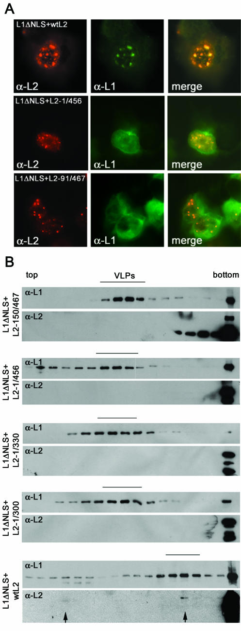
Recent reports suggest that nuclear domain(s) 10 (ND10) is the site of papillomavirus morphogenesis. The viral genome replicates in or close to ND10. In addition, the minor capsid protein, L2, accumulates in these subnuclear structures and recruits the major capsid protein, L1. We have now used cell lines deficient for promyelocytic leukemia (PML) protein, the main structural component of ND10, to study the role of this nuclear protein for L2 incorporation into virus-like particles (VLPs). L2 expressed in PML protein knockout (PML(-/-)) cells accumulated in nuclear dots, which resemble L2 aggregates forming at ND10 in PML protein-containing cells. These L2 assemblies also attracted L1 and the transcriptional repressor Daxx, suggesting that they are functional in the absence of PML protein. In addition, L2-containing VLPs assembled in PML(-/-) cells. In order to analyze whether incorporation of L2 into VLPs requires any specific subcellular localization, an L1 mutant defective for nuclear transport and L2 mutants deficient in nuclear translocation and/or ND10 localization were constructed. Using this approach, we identified two independent L2 domains interacting with L1. Mutant L2 proteins not accumulating in ND10 were incorporated into VLPs. Mutant L1 protein, which assembled into VLPs in the cytoplasm, did not incorporate L2 defective for nuclear translocation. The same mutant L2 protein, which passively diffuses into the nucleus, is incorporated into wild-type L1-VLPs in the nucleus. Our data demonstrate that the incorporation of L2 into VLPs requires nuclear but not ND10 localization.


Figures

FIG. 1.
FIG. 1.
L2 forms nuclear aggregates in PML protein knockout cells and attracts Daxx into these structures. PEF-T and PML−/− cells were grown on coverslips and subsequently infected for the indicated periods of time with VTF7-3 and vac33L2 (A and C) or with VTF7-3 and vac33L1 (D). Cells were stained for L2 alone (A), for L2 and Daxx (C), and for L1 and Daxx (D). (B) Uninfected PEF-T and PML−/− cells stained for Daxx served as controls.
FIG. 2.
FIG. 2.
L1 protein is attracted into L2 nuclear aggregates in PML−/− cells. (A) PML−/− cells were grown on coverslips and subsequently coinfected for the indicated periods of time with VTF7-3, vac33L1, and vac33L2. Cells were fixed and stained for L1 and L2. (B) Nuclear extracts were prepared from PEF-T and PML−/− cells expressing L2 alone or both L1 and L2 and then subjected to buoyant cesium chloride density gradient centrifugation. Aliquots of the VLP-containing fractions (L1 and L2) and the fraction with corresponding density (L2 alone) were analyzed by sedimentation through linear sucrose gradients. The gradients were fractionated and analyzed by L1- and L2-specific Western blot. Twelvefold more protein was used for the detection of L2 compared to L1. Cosedimentation of L1 and L2 is indicative of L2 incorporation into VLPs.
FIG. 3.
FIG. 3.
Electron micrographs of VLPs. (A) wt VLPs generated in PML−/− or PEF-T cells. (B) Mutant VLPs composed of L1ΔNLS generated in HuTK cells. Bar, 100 nm.
FIG. 4.
FIG. 4.
Two L2 domains independently interact with L1 yielding L1/L2-VLPs. (A and C) Partially purified VLPs extracted from PML−/− cells expressing L2-1/456, -1/330, -1/300 (A), or -150/467 (C), together with wt L1, were subjected to sedimentation through linear sucrose gradients. Fractions were analyzed for presence of L1 and L2 by Western blot. Fourfold more protein was used for the detection of L2 compared to L1. Inserts show the subcellular localization of the indicated L2 mutant proteins 6 h (L2-1/456, -1/330, -1/300, and -150/467) and 12 h (L2-1/300) after infection. The result for L2-1/300 at 12 h postinfection is shown to demonstrate diffusion into the nucleus. (B and D) The indicated L2 mutants were expressed together with wt L1 in PML−/− cells by using recombinant vaccinia viruses. Cells were then immunostained for L1 and L2.
FIG. 5.
FIG. 5.
wt L2 but not L2 mutant protein rescues L1ΔNLS for nuclear translocation and is incorporated into mutant VLPs. (A) The indicated L2 variants were coexpressed with L1ΔNLS in PML−/− cells for 12 h by using recombinant vaccinia viruses. L1 and L2 were visualized by immunofluorescence. (B) Incorporation of wt and mutant L2 proteins into VLPs composed of L1ΔNLS was analyzed by sedimentation through linear sucrose gradients (20 to 40% sucrose for wt L2 and 10 to 60% sucrose for mutant L2 protein) and Western blot. Fourfold more protein was used for the detection of L2 than for L1. With the exception of wt L2 no cosedimentation with L1ΔNLS was observed.

References

    1. Adamson, A. L., and S. Kenney. 2001. Epstein-barr virus immediate-early protein BZLF1 is SUMO-1 modified and disrupts promyelocytic leukemia bodies. J. Virol. 75:2388-2399. - PMC - PubMed
    1. Ahn, J. H., and G. S. Hayward. 1997. The major immediate-early proteins IE1 and IE2 of human cytomegalovirus colocalize with and disrupt PML-associated nuclear bodies at very early times in infected permissive cells. J. Virol. 71:4599-4613. - PMC - PubMed
    1. Baker, T. S., W. W. Newcomb, N. H. Olson, L. M. Cowsert, C. Olson, and J. C. Brown. 1991. Structures of bovine and human papillomaviruses: analysis by cryoelectron microscopy and three-dimensional image reconstruction. Biophys. J. 60:1445-1456. - PMC - PubMed
    1. Becker, K. A., L. Florin, C. Sapp, and M. Sapp. 2003. Dissection of human papillomavirus type 33 L2 domains involved in nuclear domain (ND) 10 homing and reorganization. Virology 314:161-167. - PubMed
    1. Day, P. M., R. B. Roden, D. R. Lowy, and J. T. Schiller. 1998. The papillomavirus minor capsid protein, L2, induces localization of the major capsid protein, L1, and the viral transcription/replication protein, E2, to PML oncogenic domains. J. Virol. 72:142-150. - PMC - PubMed

Publication types

MeSH terms

LinkOut - more resources