. 2004 Feb;78(3):1181-94.
doi: 10.1128/jvi.78.3.1181-1194.2004.

The immediate-early 63 protein of Varicella-Zoster virus: analysis of functional domains required for replication in vitro and for T-cell and skin tropism in the SCIDhu model in vivo

Affiliations

The immediate-early 63 protein of Varicella-Zoster virus: analysis of functional domains required for replication in vitro and for T-cell and skin tropism in the SCIDhu model in vivo

Armin Baiker et al. J Virol. 2004 Feb.

Abstract

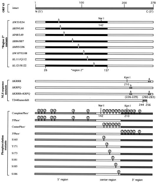
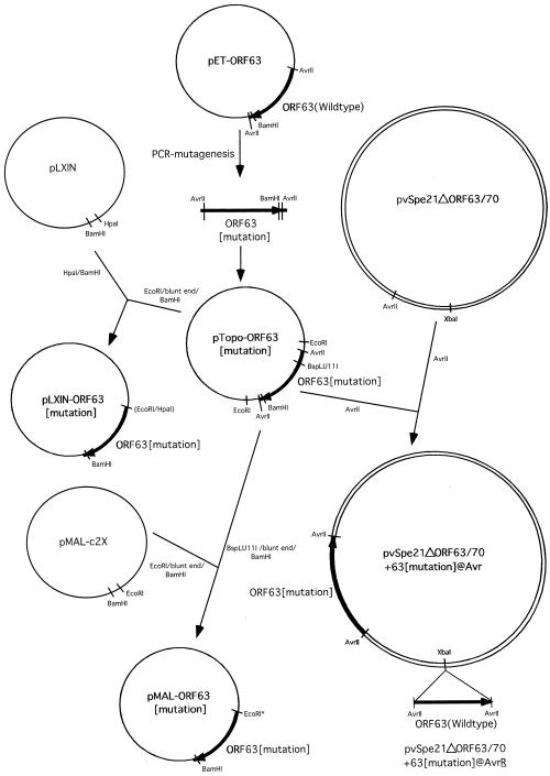
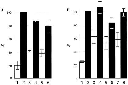
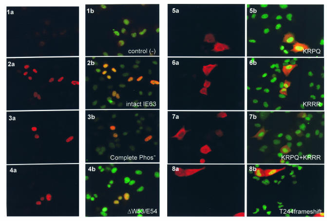
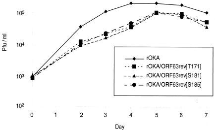
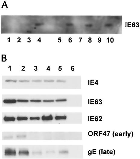
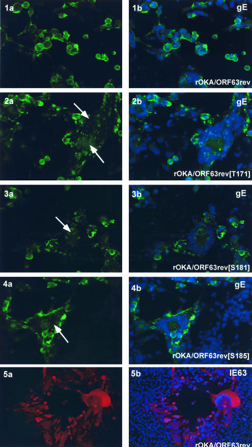
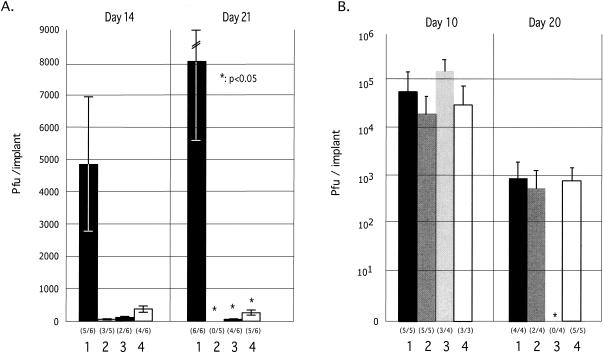
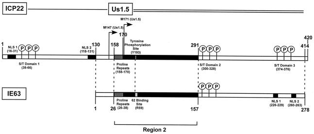
The immediate-early 63-kDa (IE63) protein of varicella-zoster virus (VZV) is a phosphoprotein encoded by open reading frame (ORF) ORF63/ORF70. To identify functional domains, 22 ORF63 mutations were evaluated for effects on IE63 binding to the major VZV transactivator, IE62, and on IE63 phosphorylation and nuclear localization in transient transfections, and after insertion into the viral genome with VZV cosmids. The IE62 binding site was mapped to IE63 amino acids 55 to 67, with R59/L60 being critical residues. Alanine substitutions within the IE63 center region showed that S165, S173, and S185 were phosphorylated by cellular kinases. Four mutations that changed two putative nuclear localization signal (NLS) sequences altered IE63 distribution to a cytoplasmic/nuclear pattern. Only three of 22 mutations in ORF63 were compatible with recovery of infectious VZV from our cosmids, but infectivity was restored by inserting intact ORF63 into each mutated cosmid. The viable IE63 mutants had a single alanine substitution, altering T171, S181, or S185. These mutants, rOKA/ORF63rev[T171], rOKA/ORF63rev[S181], and rOKA/ORF63rev[S185], produced less infectious virus and had a decreased plaque phenotype in vitro. ORF47 kinase protein and glycoprotein E (gE) synthesis was reduced, indicating that IE63 contributed to optimal expression of early and late gene products. The three IE63 mutants replicated in skin xenografts in the SCIDhu mouse model, but virulence was markedly attenuated. In contrast, infectivity in T-cell xenografts was not altered. Comparative analysis suggested that IE63 resembled the herpes simplex virus type 1 U(S)1.5 protein, which is expressed colinearly with ICP22 (U(S)1). In summary, most mutations of ORF63 made with our VZV cosmid system were lethal for infectivity. The few IE63 changes that were tolerated resulted in VZV mutants with an impaired capacity to replicate in vitro. However, the IE63 mutants were attenuated in skin but not T cells in vivo, indicating that the contribution of the IE63 tegument/regulatory protein to VZV pathogenesis depends upon the differentiated human cell type which is targeted for infection within the intact tissue microenvironment.


Figures

FIG. 1.
FIG. 1.
Overview of the mutational analysis of IE63 protein. This figure depicts all of the amino acid changes that were introduced into IE63 protein. Mutations in the IE63 region 2 deleted the conserved amino acid pairs ΔW53/E54, ΔR59/L60, ΔF68/L69, ΔR86/R87, ΔM95/G96, ΔW107/E108, ΔL111/Q112, and ΔL121/R122. Nuclear localization sequence mutations deleted the four amino acids 226 to 229 (ΔKRPQ) or 260 to 263 (ΔKRRR), by removing both putative NLS sequences (ΔKRRR+KRPQ) and by creating a frameshift, replacing the full-length 278-amino-acid protein with an IE63 truncation at amino acid 256 (T244 frameshift). Mutations of putative phosphorylation sites were made by alanine substitution of 19 serine (S) or threonine (T) residues that were predicted targets of cellular S/T kinases, including S12, S13, S15, T41, S42, S82, S129, S165, T171, S173, S181, S185, S186, S197, T201, S203, T222, S224, and T244 (CompletePhos mutant). The mutant 5′Phos contains alanine substitutions of the first seven putative phosphorylation targets (S12 to S129) in the IE63 N terminus, the mutant CenterPhos has substitutions of the six putative phosphorylation sites in the center region (S165 to S186), and the mutant 3′Phos has substitutions of the six putative phosphorylation sites (S197 to T244) in the IE63 C terminus. Single alanine substitutions were made in the S and T residues S165, T171, S173, S181, S185, and S186 in the center segment of the IE63 protein.
FIG. 2.
FIG. 2.
Construction of ORF63 mutations. This figure is a schematic representation of the cloning steps involved in the construction of all plasmids and cosmids that were used to make ORF63 mutations, as described in Materials and Methods.
FIG. 3.
FIG. 3.
Mapping of the IE62 binding site in IE63 protein. The IE62 pull-down assay was performed with IE63-MBP fusion proteins that had deletions of the conserved amino acid pairs in region 2. The ORF63 mutations were made in pMAL-c2X and expressed in E. coli (BL21-AI) as fusion proteins at the MBP C terminus. The recombinant proteins were affinity purified with amylose resin (New England Biolabs, Beverly, Mass.) and incubated with recombinant IE62. IE62 that bound to the IE63 recombinant proteins was detected with polyclonal rabbit anti-ORF62 antibody. IE62 binding is shown to intact IE63 (lane 1), the IE63 mutants ΔW53/E54 (lane 3), ΔF68/L69 (lane 5), ΔR86/R87 (lane 6), ΔM95/G96 (lane 7), ΔW107/E108 (lane 8), ΔL111/Q112 (lane 9), ΔL121/R122 (lane 10), and 5′Phos (lane 11). No IE62 was recovered in the pull-down assay done with MBP alone (lane 2) or with the IE63-MBP fusion protein ΔR59/L60 (lane 4).
FIG. 4.
FIG. 4.
Kinase assay to evaluate phosphorylation of IE63 mutants expressed in 293T cells. 293T cells were transfected with the pLXIN-based ORF63 mutants designed to substitute alanine for S or T residues that were putative phosphorylation targets, or with control plasmids, and harvested in RIPA buffer after 36 h. IE63 was immunoprecipitated with polyclonal rabbit antiserum, and IE63 phosphorylation by cellular S/T kinases was assessed with radiolabeled ATP, followed by SDS gel electrophoresis and phosphorimager detection. The bar graph in panel A shows the results of three independent kinase assays performed with the multiple putative phosphorylation site mutants and reported as a mean percentage relative to intact IE63 (bar 2). The other bars are 1, negative control (pLXIN), 3, CompletePhos mutant, 4, 5′Phos mutant, 5, CenterPhos mutant; and 6, 3′Phos mutant. Panel B shows the combined results of three independent kinase assays performed with the mutants disrupting single putative phosphorylation targets relative to intact IE63 (no. 2); the other bars are 1, negative control (pLXIN), 3, S165, 4, T171, 5, S173, 6, S181, 7, S185; and 8, S186. The lines indicate standard errors.
FIG. 5.
FIG. 5.
Effects of IE63 mutations in putative nuclear localization signal sequences. Intracellular distribution of the IE63 mutant proteins expressed as pLXIN constructs in transiently transfected HeLa cells was examined by immunofluorescence microscopy 48 h after transfection, with polyclonal rabbit antiserum against IE63 (indocarbocyanine label: red; panels 1a to 8a) and dual stain with anti-ORF63 and anti-human nuclei, a murine monoclonal antibody (fluorescein isothiocyanate label: green; panels 1b to 8b). The constructs tested were plasmid alone (1a, 1b), intact IE63 (2a, 2b), CompletePhos- (3a, 3b), ΔW53/W54 (4a, 4b), ΔKRPQ (5a, 5b), ΔKRRR (6a, 6b), ΔKRPQ+ΔKRRR (7a, 7b), and T244 frameshift (8a, 8b). Magnification: 40×.
FIG. 6.
FIG. 6.
Replication of IE63 mutant viruses in melanoma cells. Melanoma cells were inoculated on day 0 with 103 PFU of rOka or rOka mutants rOKA/ORF63rev[T171], rOKA/ORF63rev[S181], and rOKA/ORF63rev[S185]. Aliquots were harvested for 7 days, and infectious foci were determined by titration on melanoma cell monolayers. Each point represents the mean of three wells.
FIG. 7.
FIG. 7.
Kinase assay to evaluate phosphorylation of IE63 as expressed in IE63 mutant viruses. IE63 was immunoprecipitated with polyclonal rabbit antiserum and phosphorylation was monitored by incubation of the immunoprecipitate with radiolabeled ATP, followed by SDS gel electrophoresis and phosphorimager detection. Panel A shows phosphorylation of IE63 in cells infected with rOka (bar 2) or rOKAORF47ΔC, an ORF47 kinase null mutant (bar 3), and uninfected melanoma cell control (bar 1). The bar graph in panel B shows the results of two independent kinase assays performed with IE63 mutant viruses and reported as a mean percentage relative to cells infected with rOKA/ORF63rev (bar 2); the other bars are uninfected melanoma cell control (bar 1), rOKA/ORF63rev[T171] (bar 3), rOKA/ORF63rev[S181] (bar 4), and rOKA/ORF63rev[S185] (bar 5). The lines indicate standard errors.
FIG. 8.
FIG. 8.
Immunoprecipitation of IE62 protein from melanoma cells infected with IE63 mutant viruses with anti-IE63 antiserum and expression of VZV immediate-early, early, and late proteins by IE63 mutant viruses. As shown in panel A, IE62 in lysates of infected cells was immunoprecipitated with monoclonal antibody against IE62. IE63 that coimmunoprecipitated with IE62 was detected with polyclonal rabbit antiserum against IE63 (even-numbered lanes) or nonspecific murine IgG (odd-numbered lanes). Samples tested included lanes 1 and 2, uninfected melanoma cells; lanes 3 and 4; rOKA/ORF63rev-infected cells; lanes 5 and 6, rOKA/ORF63rev[T171]-infected cells; lanes 7 and 8, rOKA/ORF63rev[S181]-infected cells; and lanes 9 and 10, rOKA/ORF63rev[S185]-infected cells. As shown in panel B, melanoma cells were infected with 5 × 10⁴ PFU of the test virus and harvested at 4 days postinfection. Cell lysates were evaluated for expression of immediate-early proteins IE62 and IE63 and ORF4 proteins, the ORF47 viral kinase, which is an early protein, and the late protein gE, with rabbit polyclonal antiserum specific for IE62, IE63, ORF4, or ORF47, and a murine monoclonal antibody to gE. The specimens tested were lane 1, rOka (contains ORFs 63 and 70); lane 2, rOKA/ORF63rev (one copy of ORF63 at a nonnative site); lane 3, rOKA/ORF63rev[T171]; lane 4, rOKA/ORF63rev[S181]; and lane 5, rOKA/ORF63rev[S185].
FIG. 9.
FIG. 9.
Intracellular localization of VZV IE63 and gE in cells infected with IE63 mutant viruses. Melanoma cells were infected with rOKA/ORF63rev (1 a,b; 5 a,b), rOKA/ORF63rev[T171] (2 a,b), rOKA/ORF63rev[T181] (3 a.b), or rOKA/ORF63rev[S185] (4 a, b). Monolayers were stained with anti-rabbit IE63 antiserum (red) and counterstained with the nuclear marker, 4′,6′-diamidino-2-phenylindole (DAPI) (blue) (panel 5 a, b) or with anti-gE monoclonal antibody (fluorescein isothiocyanate: green) (panels 1 to 4) and counterstained with the nuclear marker DAPI (blue). Panels 2a to 4a, arrows indicate abnormal, irregular polykaryocytes with punctate distribution of gE. Immunofluorescence microscopy was performed at 4 days after infection (late). Magnification, 40×.
FIG. 10.
FIG. 10.
Replication of IE63 mutant viruses in skin and T-cell xenografts in SCID-hu mice. Skin xenografts in SCID mice were injected with (left to right) rOKA, rOKA/ORF63rev[T171], rOKA/ORF63rev[S181], or rOKA/ORF63rev[S185] having equivalent inoculum titers. Virus titers in skin xenografts were assessed after harvest at day 14 (A, left panel) and day 21 (A, right panel) after inoculation and were graphed as mean titers for xenografts that yielded infectious virus, with lines indicating standard errors. The number of xenografts from which infectious virus was recovered per number that were inoculated is given in parentheses below the horizontal axis. The P values were <0.05 when titers of rOka and each of the IE63 mutant viruses were compared at day 21. Replication of VZV recombinants in T cells was assessed at days 10 and 20 (B). Lines indicate the standard errors.
FIG. 11.
FIG. 11.
Comparative analysis of IE63 and herpes simplex virus type 1 ICP22 and US1.5 gene products. This diagram illustrates the similarities and differences between IE63 and the herpes simplex virus type 1 homologue ICP22 and the colinear US1.5 gene product. The N-terminal sequence of ICP22 is not present in IE63. IE63 is similar in size to the US1.5 gene product, and both proteins have similar regions arranged in the same linear pattern, e.g., initial proline repeats, the conserved region 2, and an S/T-rich center domain. Nuclear localization signal (NLS) sequences located in the C terminus of IE63 are not present in US1.5; US1.5 has another S/T domain where the NLS domains appear in IE63, and US1.5 has six amino acid residues at the C terminus that are not found in IE63. Amino acid identity between IE63 and US1.5 is 24.9%, and homology is 46%.

References

    1. Arvin, A. M. 2001. Varicella-zoster virus, p. 2731-2767. In D. M. Knipe and P. M. Howley (ed.), Fields virology. Lippincott-Raven, Philadelphia, Pa.
    1. Bateman, A., E. Birney, R. Durbin, S. R. Eddy, K. L. Howe, and E. L. Sonnhammer. 2000. The Pfam protein families database. Nucleic Acids Res. 28:263-266. - PMC - PubMed
    1. Besser, J., M. H. Sommer, L. Zerboni, C. Bagowski, H. Ito, J. Moffat, Ku, C.-C. and A. M. Arvin. 2003. Differentiation of varicella-zoster virus ORF47 protein kinase and IE62 protein binding domains and their contributions to replication in human skin xenografts in the SCIDhu mouse. J. Virol. 77:5964-5974. - PMC - PubMed
    1. Blom, N., S. Gammeltoft, und S. Brunak. 1999. Sequence and structure-based prediction of eukaryotic protein phosphorylation sites. J. Mol. Biol. 294:1351-1362. - PubMed
    1. Bontems, S., Valentin. D., E L. Baudox, B. Rentier, Sadzot-Delvaux, C., and J. Piette. 2002. Phosphorylation of varicella-zoster virus IE63 protein by casein kinases influences its cellular localization and gene regulation activity. J. Biol. Chem. 277:21050-21060. - PubMed

Publication types

MeSH terms

Substances

LinkOut - more resources