. 2004 Feb;78(3):1375-83.
doi: 10.1128/jvi.78.3.1375-1383.2004.

Compensatory link between fusion and endocytosis of human immunodeficiency virus type 1 in human CD4 T lymphocytes

Affiliations

Compensatory link between fusion and endocytosis of human immunodeficiency virus type 1 in human CD4 T lymphocytes

Evelyne Schaeffer et al. J Virol. 2004 Feb.

Abstract

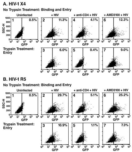
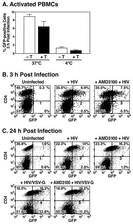
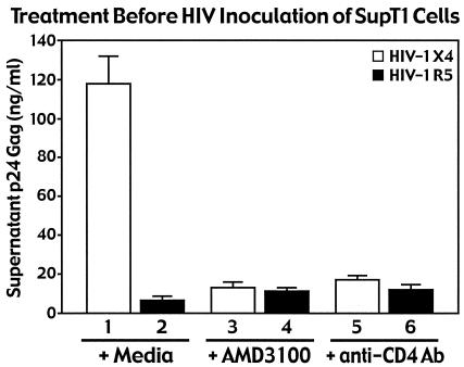
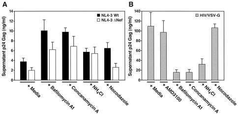
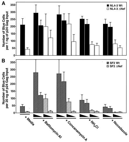
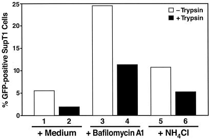
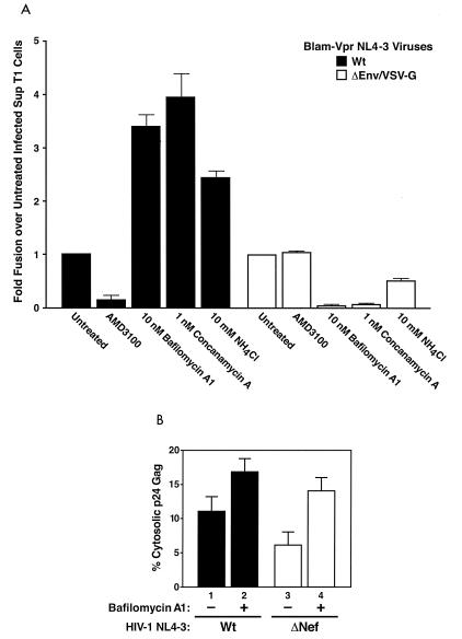
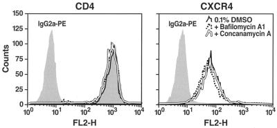
Virions of the type 1 human immunodeficiency virus (HIV-1) can enter target cells by fusion or endocytosis, with sharply different functional consequences. Fusion promotes productive infection of the target cell, while endocytosis generally leads to virion inactivation in acidified endosomes or degradation in lysosomes. Virion fusion and endocytosis occur equally in T cells, but these pathways have been regarded as independent because endocytosis of HIV virions requires neither CD4 nor CCR5/CXCR4 engagement in HeLa-CD4 cells. Using flow cytometric techniques to assess the binding and entry of green fluorescent protein (GFP)-Vpr-labeled HIV virions into primary peripheral blood mononuclear cells, we have found that HIV fusion and endocytosis are restricted to the CD4-expressing subset of cells and that both pathways commonly require the initial binding of HIV virions to surface CD4 receptors. Blockade of CXCR4-tropic HIV virion fusion with AMD3100, a CXCR4-specific entry inhibitor, increased virion entry via the endocytic pathway. Similarly, inhibition of endosome acidification with bafilomycin A1, concanamycin A, or NH(4)Cl enhanced entry via the fusion pathway. Although fusion remained dependent on CD4 and chemokine receptor binding, the endosome inhibitors did not alter surface expression of CD4 and CXCR4. These results suggest that fusion in the presence of the endosome inhibitors likely occurs within nonacidified endosomes. However, the ability of these inhibitors to impair vesicle trafficking from early to late endosomes in some cells could also increase the recycling of these virion-containing endosomes to the cell surface, where fusion occurs. In summary, our results reveal an unexpected, CD4-mediated reciprocal relationship between the pathways governing HIV virion fusion and endocytosis.


Figures

FIG. 1.
FIG. 1.
Flow cytometric analysis of human SupT1 cells incubated with GFP-Vpr-labeled X4-tropic (A) or R5-tropic (B) HIV-1 virions in the presence of medium (panels 2 and 3), neutralizing anti-CD4 antibodies (panels 4 and 5), or AMD3100 (panels 6 and 7). SupT1 T cells were preincubated in the presence or absence of anti-CD4 antibodies or AMD3100 for 30 min at 37°C. Cells were then inoculated with GFP-Vpr-labeled X4-tropic HIV-1 or CCR5-tropic HIV-1 R5 (200 ng of p24 Gag) for 3 h at 37°C. The cells were subsequently washed with PBS (panels 1, 2, 4, and 6) or treated with a trypsin-EDTA solution (panels 3, 5, and 7) to remove surface-bound virions. Live cells were then analyzed by flow cytometry for GFP epifluorescence. The vertical bar indicates a gate established by analysis of uninfected cells (panels 1). The percentage of GFP-positive cells is indicated in the upper right hand corner of each panel. The anti-CD4 antibodies significantly inhibited the entry of both X4-tropic and R5-tropic virions, whereas AMD3100 unexpectedly failed to reduce the overall entry of X4-tropic virions.
FIG. 2.
FIG. 2.
Flow cytometric analysis of PHA-activated human lymphoblasts infected with GFP-Vpr-labeled HIV-1 virions. (A) PBMCs activated with PHA and cultured in interleukin-2 for 4 days were inoculated with GFP-Vpr-labeled X4-tropic HIV-1 (300 ng of p24 Gag) at 37 or 4°C as indicated. After 3 h, the cells were washed at room temperature with PBS (−T) or a trypsin-EDTA solution (+T) to remove surface-bound virions. Live cells were then analyzed by flow cytometry to detect GFP epifluorescence. Error bars indicate standard deviations of the mean derived from two independent experiments. (B and C) PHA-activated human lymphoblasts were pretreated for 30 min at 37°C with either medium (panels 2 and 4) or AMD3100 (panels 3 and 5). Cells were then incubated at 37°C for 3 or 24 h with GFP-Vpr-labeled X4-tropic HIV-1 or HIV virions pseudotyped with the VSV-G envelope (200 ng of p24 Gag) at 37°C. Cells were subsequently stained with APC-conjugated CD4 antibodies (B) or PE-conjugated CD4 antibodies (C). Cells were analyzed for GFP epifluorescence and APC or PE immunofluorescence. The percentage of cells in each quadrant is indicated. Data shown are from a representative experiment performed three times with comparable results. Note preferential entry of HIV into CD4 cells and the absence of inhibitory effects of AMD3100 measured either at 3 or 24 h.
FIG. 3.
FIG. 3.
Neutralizing anti-CD4 antibodies and the X4-specific AMD3100 entry inhibitor efficiently block replication of X4-tropic HIV in SupT1 cells. HIV-1 NL4-3 X4 and R5 viruses (100 ng of p24 Gag) were incubated with SupT1 cells pretreated for 30 min at 37°C with AMD3100 or neutralizing anti-CD4 antibodies. After 3 h at 37°C, the cultures were extensively washed. After 2 days of culture, p24 Gag present in the cell medium was quantitated by ELISA. Data represent an average of three independent infections performed in duplicate. Error bars indicate standard deviations. AMD3100 did not impair overall entry of X4-tropic virions into SupT1 cells (Fig. 1A) but markedly impaired viral replication.
FIG. 4.
FIG. 4.
Inhibitors of endosome acidification enhance HIV-1 replication but impair HIV-1/VSV-G pseudotype replication in SupT1 T cells. SupT1 cells were preincubated for 30 min at 37°C with DMSO (0.1%; diluent control), bafilomycin A1 (100 nM), concanamycin A (0.6 nM), NH4Cl (10 mM), or nocodazole (1 μM). Cells were infected with X4-tropic HIV WT (WT, closed bars) or HIVΔNef (open bars) (A) or HIV virions pseudotyped with the VSV-G envelope (50 ng of p24 Gag) (B). After incubation for 3 h at 37°C, the cells were washed. Two days later, p24 Gag in the culture medium was quantitated by ELISA. Data represent an average for three independent infections performed in duplicate. Error bars indicate standard deviations. The endosome inhibitors bafilomycin A1, concanamycin A, and NH4Cl enhanced the replication of both WT and ΔNef viruses but impaired replication of VSV-G pseudotypes of HIV where fusion is dependent on endosome acidification.
FIG. 5.
FIG. 5.
Endosome inhibitors enhance HIV-1 replication in MAGI cells. (A and B) MAGI cells were pretreated for 45 min at 37°C with DMSO (0.1%; diluent control), bafilomycin A1 (10 and 1 nM), concanamycin A (1.2 and 0.6 nM), NH4Cl (10 and 1 mM), or nocodazole (200 and 20 nM) and inoculated with HIV-1 NL4-3 WT or ΔNef viruses (1 ng of p24 Gag) (A) or HIV-SF2 WT and ΔNef viruses (20 ng of p24 Gag) (B) as indicated. Foci of blue cells reflecting Tat activation of the integrated HIV-LTR-β-galactosidase reporter gene were enumerated 48 h later. Values are representative of at least three independent experiments performed in duplicate. Error bars indicate standard deviations The endosome inhibitors enhanced the infectivity of WT HIV, but the greatest effects were obtained with viruses displaying intrinsically diminished infectivity (SF2 and ΔNef).
FIG. 6.
FIG. 6.
Both surface binding and intracellular entry by fusion of HIV-1 virions are enhanced by endosome inhibitors. SupT1 cells were pretreated for 30 min at 37°C with medium (columns 1 and 2), bafilomycin A1 (100 nM; columns 3 and 4), or NH4Cl (10 mM; columns 5 and 6). Cells were then incubated with GFP-Vpr-labeled HIV-1 virions (200 ng of p24 Gag) for 3 h at 37°C. Cells were either washed in PBS (columns 1, 3, and 5) or treated with a trypsin-EDTA solution to remove surface-bound virions (columns 2, 4, and 6) followed by flow cytometric analysis of living cells. The percentage of SupT1 cells displaying GFP epifluorescence is indicated on the ordinate. The data presented are from a representative experiment repeated three times with comparable results.
FIG. 7.
FIG. 7.
(A) Endosome inhibitors enhance HIV-1 fusion in SupT1 cells. SupT1 cells were pretreated for 1 h at 37°C with DMSO (0.1%; diluent control), AMD3100 (250 nM), bafilomycin A1 (10 nM), concanamycin A (1 nM), or NH4Cl (10 mM) and inoculated with BlaM-Vpr-containing viruses, either WT NL4-3 or VSV-G-pseudotyped NL4-3Δenv (25 ng or 100 ng of p24 Gag), as indicated, for 3 h. The fusion assay was performed as described in Materials and Methods, and the percentage of blue cells was determined. Shown is the fold increase in fusion, which is the ratio of percent blue cells upon treatment with indicated drug to percent blue cells without any treatment for the given virus. Thus, entry of virus into untreated cells is equal to 1. The values are representative of at least three independent experiments performed in triplicate. Error bars are indicated. The endosome inhibitors enhanced the entry of WT NL4-3 viruses while inhibiting the entry of the VSV-G-pseudotyped NL4-3Δenv viruses. In contrast, AMD3100 treatment inhibited entry of WT NL4-3 virus only and not the VSV-G-pseudotyped virus. (B) Cytosolic entry of HIV-1 particles is enhanced by bafilomycin A1. MAGI cells were treated with 0.1% DMS0 (diluent control) or bafilomycin A1 (20 nM) and infected with the indicated HIV-1-NL4-3 WT or HIV-NL4-3 ΔNef viruses (500 ng of p24 Gag). After a 1-h incubation of cells and viruses at 37°C, cytosolic fractions were prepared as described elsewhere (30). The level of p24 Gag present in each fraction was quantitated by ELISA. Values correspond to the percentage of p24 Gag present in the cytosolic fraction relative to the total intracellular p24 Gag. The total intracellular p24 Gag was approximately 1,000 pg, representing 0.2% of the input. Data are representative of seven experiments performed in duplicate. Error bars indicate standard deviations. Bafilomycin A1 enhanced cytoplasmic delivery of p24 Gag with both viruses, indicating increased fusion; however, greater relative effects were obtained with the less infectious HIV ΔNef virus.
FIG. 8.
FIG. 8.
Bafilomycin A1 and concanamycin A treatment do not alter cell surface expression of either CD4 or CXCR4. SupT1 cells were incubated for 3 h at 37°C with medium containing or lacking 0.1% DMSO, bafilomycin A1 (100 nM), or concanamycin A (0.6 nM). Cells were then washed and immunostained at 4°C with PE-conjugated anti-CD4, anti-CXCR4, or isotype control IgG2a monoclonal antibodies. Immunofluorescence intensities were analyzed by flow cytometry.

References

    1. Aiken, C. 1997. Pseudotyping human immunodeficiency virus type 1 (HIV-1) by the glycoprotein of vesicular stomatitis virus targets HIV-1 entry to an endocytic pathway and suppresses both the requirement for Nef and the sensitivity to cyclosporin A. J. Virol. 71:5871-5877. - PMC - PubMed
    1. Bayer, N., D. Schober, E. Prchla, R. F. Murphy, D. Blaas, and R. Fuchs. 1998. Effect of bafilomycin A1 and nocodazole on endocytic transport in HeLa cells: implications for viral uncoating and infection. J. Virol. 72:9645-9655. - PMC - PubMed
    1. Binley, J., and J. P. Moore. 1997. HIV-cell fusion. The viral mousetrap. Nature 387:346-348. - PubMed
    1. Blauvelt, A., H. Asada, M. W. Saville, V. Klaus-Kovtun, D. J. Altman, R. Yarchoan, and S. I. Katz. 1997. Productive infection of dendritic cells by HIV-1 and their ability to capture virus are mediated through separate pathways. J. Clin. Investig. 100:2043-2053. - PMC - PubMed
    1. Bridger, G. J., R. T. Skerlj, D. Thornton, S. Padmanabhan, S. A. Martellucci, G. W. Henson, M. J. Abrams, N. Yamamoto, K. De Vreese, R. Pauwels, et al. 1995. Synthesis and structure-activity relationships of phenylenebis(methylene)-linked bis-tetraazamacrocycles that inhibit HIV replication. Effects of macrocyclic ring size and substituents on the aromatic linker. J. Med. Chem. 38:366-378. - PubMed

Publication types

Substances