. 2004 Feb;78(3):1540-51.
doi: 10.1128/jvi.78.3.1540-1551.2004.

Effects of mutations in the cytoplasmic domain of herpes simplex virus type 1 glycoprotein B on intracellular transport and infectivity

Affiliations

Effects of mutations in the cytoplasmic domain of herpes simplex virus type 1 glycoprotein B on intracellular transport and infectivity

Igor Beitia Ortiz de Zarate et al. J Virol. 2004 Feb.

Abstract

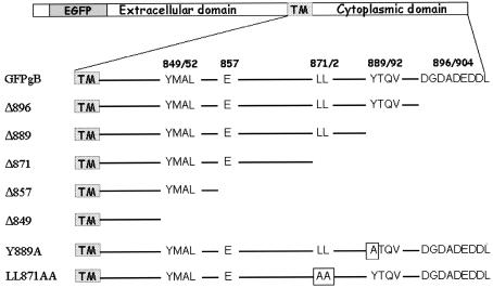
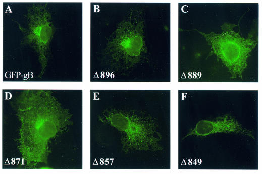
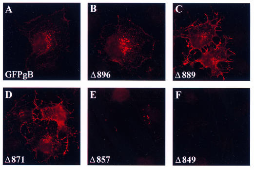
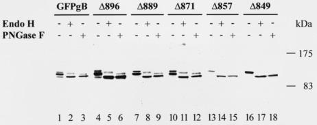
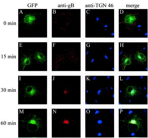
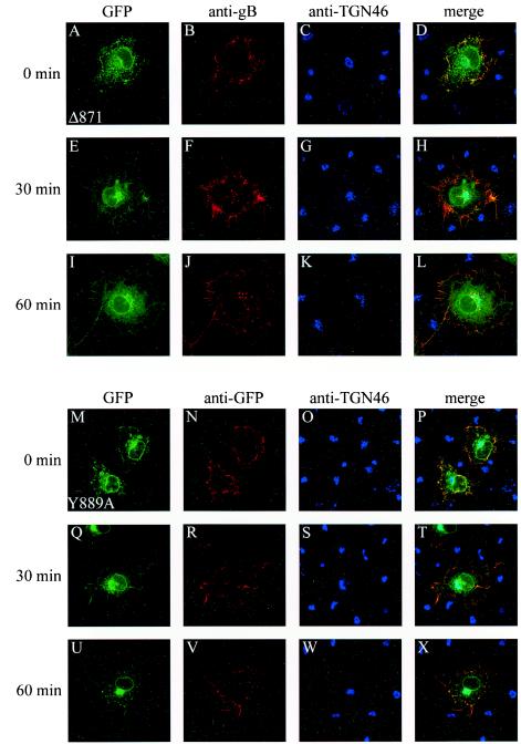
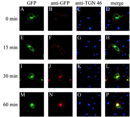
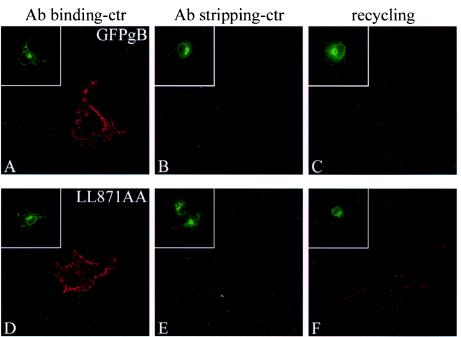
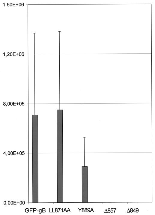
Herpes simplex virus type 1 (HSV-1) is a human pathogen of the alphaherpesvirus family which infects and spreads in the nervous system. Glycoproteins play a key role in the process of assembly and maturation of herpesviruses, which is essential for neuroinvasion and transneuronal spread. Glycoprotein B (gB) is a main component of the HSV-1 envelope and is necessary for the production of infectious particles. The cytoplasmic domain of gB, the longest one among HSV-1 glycoproteins, contains several highly conserved peptide sequences homologous to motifs involved in intracellular sorting. To determine the specific roles of these motifs in processing, subcellular localization, and the capacity of HSV-1 gB to complement a gB-null virus, we generated truncated or point mutated forms of a green fluorescent protein (GFP)-tagged gB. GFP-gB with a deletion in the acidic cluster DGDADEDDL (amino acids [aa] 896 to 904) behaved the same as the parental form. Deletion or disruption of the YTQV motif (aa 889 to 892) abolished internalization and reduced complementation by 60%. Disruption of the LL motif (aa 871 to 872) impaired the return of the protein to the trans-Golgi network (TGN) while enhancing its recycling to the plasma membrane. Truncations from residue E 857 abolished transport and processing of the truncated proteins, which had null complementation activity, through the Golgi complex. Altogether, our results favor a model in which HSV-1 gets its final envelope in the TGN, and they suggest that endocytosis, albeit not necessary, might play a role in infectivity.


Figures

FIG. 1.
FIG. 1.
Representation of HSV-1 gB cytoplasmic domain. The top drawing depicts the EGFP-tagged gB. The line below depicts the cytoplasmic domain and the locations of highly conserved sorting motifs. The amino acid numbers bracketing the motifs are shown above. The bottom seven lines show the cytoplasmic domains of the mutated gB forms used in the study. Altered amino acids are shown in shaded boxes.
FIG. 2.
FIG. 2.
Comparative localization of GFP-gB and the truncated forms in transfected cells. Cos-7 cells were transfected with pGFP-gB or each of the truncated forms. Images show spontaneous GFP fluorescence 24 h after transfection. Fluorescence was visualized with a Zeiss Axiophot conventional microscope.
FIG. 3.
FIG. 3.
Surface labeling of GFP-gB and the truncated forms in transfected cells. Cos-7 cells were fixed under nonpermeabilizing conditions 24 h after transfection with pGFP-gB or each of the truncated forms and were labeled with an anti-gB antibody and a Texas red-labeled secondary antibody. Fluorescence was visualized with a Zeiss Axiophot conventional microscope.
FIG. 4.
FIG. 4.
Immunoblot analysis. Cos-7 cells were transfected with pGFP-gB or each of the truncated forms and were harvested 48 h later. Lysates were treated with endo H or PNGase F, as indicated above each lane, and subjected to denaturing sodium dodecyl sulfate-polyacrylamide gel electrophoresis. Immunoblots were prepared and probed with an anti-GFP antibody.
FIG. 5.
FIG. 5.
Internalization of GFP-gB. Cos-7 cells expressing GFP-gB were incubated at 4°C with an anti-gB antibody for 1 h and then returned to 37°C for 0 min (A to D), 15 min (E to H), 30 min (I to L), or 60 min (M to P). Cells were fixed in methanol and labeled with an anti-TGN 46 antibody. Images show spontaneous GFP fluorescence (A, E, I, and M) and indirect immunofluorescence from the anti-gB antibody and a Cy3-labeled secondary antibody (B, F, J, and N) or from the anti-TGN 46 antibody and Cy5-labeled secondary antibody (C, G, K, and O). (D, H, L, and P) Merged images. Fluorescence was visualized with a Bio-Rad MRC 1000 confocal microscope.
FIG.6.
FIG.6.
Internalization of Δ871 and Y889A. The same experiment as that for Fig. 5 was reproduced in Cos-7 cells expressing the truncated Δ871 protein or the mutated Y889A gB protein. Subcellular localization was examined by confocal microscopy as described in the legend for Fig. 5. (A to D and M to P) 0-min incubation at 37°C; (E to H and Q to T) 30-min incubation at 37°C; (I to L and U to X) 60-min incubation at 37°C. EGFP fluorescence of Δ871 and of Y889A is shown in panels A, E, and I and panels M, Q, and U, respectively; anti-gB staining of Δ871 and of Y889A is shown in panels B, F, and J and panels N, R, and V, respectively; and anti-TGN 46 staining of Δ871 and of Y889A is shown in panels C, G, and K and panels O, S, and W, respectively. (D, H, L, P, T, and X) Merged images.
FIG. 7.
FIG. 7.
Effect of mutation of the LL motif on internalization. After transfection, LL871AA-expressing cells were incubated with an anti-GFP monoclonal antibody for 1 h and then returned to 37°C for different times. After methanol fixation and labeling with an anti-TGN 46 antibody, cells were incubated with Cy3- and Cy5-labeled secondary antibodies. Panels A to D, E to H, I to L, and M to P correspond to 0-, 15-, 30-, and 60-min incubation times, respectively, at 37°C. (A, E, I, and M) GFP fluorescence; (B, F, J, and N) anti-GFP immunofluorescence; (C, G, K, and O) TGN 46 localization; (D, H, L, and P) merged images.
FIG. 8.
FIG. 8.
Recycling of internalized GFP-gB and LL871AA to the plasma membrane. After transfection, GFP-gB- or LL871AA-expressing cells were stained with an anti-GFP antibody at 4°C and then either immediately fixed (A and D) or incubated for 20 min at 37°C to allow internalization (B, C, E, and F). After incubation, cells were subjected to an acid-glycine treatment and then immediately fixed under nonpermeabilizing conditions (B and E) or placed again at 37°C for 30 min before fixation (C and F). Fixed cells were labeled with a Cy3-coupled secondary antibody and examined by confocal microscopy. Insets show the spontaneous GFP fluorescence emitted in transfected cells.
FIG. 9.
FIG. 9.
Complementation of K082 virus by GFP-gB or the mutated Y889A, LL871AA, Δ857, or Δ849 protein. Vero cells were transfected with plasmids expressing GFP-gB or the mutated proteins and were infected 24 h later with K082 at a multiplicity of infection of 1. Extracellular virions were washed off at 1 h postinfection by treatment with an acid-glycine-saline solution, followed by three washes with PBS. Infected cell culture supernatants were harvested 24 h after infection and titrated on D6 and Vero cells. Titers shown are the averages from three independent experiments.

Similar articles

Cited by

References

    1. Baghian, A., L. Huang, S. Newman, S. Jayachandra, and K. G. Kousoulas. 1993. Truncation of the carboxy-terminal 28 amino acids of glycoprotein B specified by herpes simplex virus type 1 mutant amb1511-7 causes extensive cell fusion. J. Virol. 67:2396-2401. - PMC - PubMed
    1. Berlioz-Torrent, C., B. L. Shacklett, L. Erdtmann, L. Delamarre, I. Bouchaert, P. Sonigo, M. C. Dokhelar, and R. Benarous. 1999. Interactions of the cytoplasmic domains of human and simian retroviral transmembrane proteins with components of the clathrin adaptor complexes modulate intracellular and cell surface expression of envelope glycoproteins. J. Virol. 73:1350-1361. - PMC - PubMed
    1. Blot, G., K. Janvier, S. Le Panse, R. Benarous, and C. Berlioz-Torrent. 2003. Targeting of the human immunodeficiency virus type 1 envelope to the trans-Golgi network through binding to TIP47 is required for env incorporation into virions and infectivity. J. Virol. 77:6931-6945. - PMC - PubMed
    1. Bonneau, R. H., L. A. Salvucci, D. C. Johnson, and S. S. Tevethia. 1993. Epitope specificity of H-2Kb-restricted, HSV-1-, and HSV-2-cross-reactive cytotoxic T lymphocyte clones. Virology 195:62-70. - PubMed
    1. Brideau, A. D., L. W. Enquist, and R. S. Tirabassi. 2000. The role of virion membrane protein endocytosis in the herpesvirus life cycle. J. Clin. Virol. 17:69-82. - PubMed

Publication types

MeSH terms

Substances

LinkOut - more resources