. 2004 Jan;113(2):188-99.
doi: 10.1172/JCI19383.

Soluble VEGF isoforms are essential for establishing epiphyseal vascularization and regulating chondrocyte development and survival

Affiliations

Soluble VEGF isoforms are essential for establishing epiphyseal vascularization and regulating chondrocyte development and survival

Christa Maes et al. J Clin Invest. 2004 Jan.

Abstract

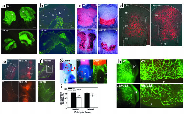
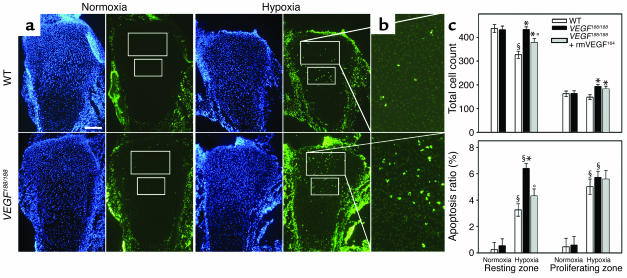
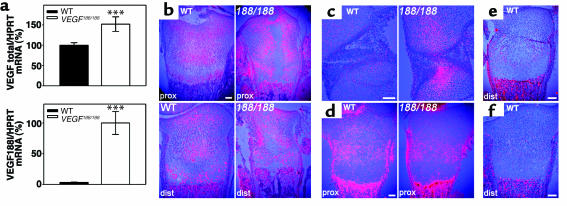
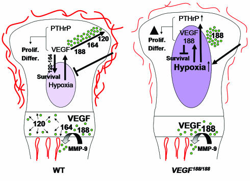
VEGF is crucial for metaphyseal bone vascularization. In contrast, the angiogenic factors required for vascularization of epiphyseal cartilage are unknown, although this represents a developmentally and clinically important aspect of bone growth. The VEGF gene is alternatively transcribed into VEGF(120), VEGF(164), and VEGF(188) isoforms that differ in matrix association and receptor binding. Their role in bone development was studied in mice expressing single isoforms. Here we report that expression of only VEGF(164) or only VEGF(188) (in VEGF(188/188) mice) was sufficient for metaphyseal development. VEGF(188/188) mice, however, showed dwarfism, disrupted development of growth plates and secondary ossification centers, and knee joint dysplasia. This phenotype was at least partly due to impaired vascularization surrounding the epiphysis, resulting in ectopically increased hypoxia and massive chondrocyte apoptosis in the interior of the epiphyseal cartilage. In addition to the vascular defect, we provide in vitro evidence that the VEGF(188) isoform alone is also insufficient to regulate chondrocyte proliferation and survival responses to hypoxia. Consistent herewith, chondrocytes in or close to the hypoxic zone in VEGF(188/188) mice showed increased proliferation and decreased differentiation. These findings indicate that the insoluble VEGF(188) isoform is insufficient for establishing epiphyseal vascularization and regulating cartilage development during endochondral bone formation.


Figures

Figure 1
Figure 1
Abnormal bone, cartilage, and joint development in VEGF188/188 mice. (a) Lateral view of WT and VEGF188/188 (188/188) embryos at E16.5. (b) Skeletal preparation of pups at P1.5. (c) Calvaria at P1.5 (top) and P5 (bottom), showing delayed bone growth in the mutants. (d) P1.5 tails, showing reduced size and ossification in VEGF188/188 mice. Asterisks indicate the most distal vertebrae with ossified center. (e) Forelimbs and (f) hind limbs at P1.5. Note incomplete ossification in talus and calcaneus (arrows and insets) and decreased length of the ossified diaphysis, but increased cartilage length in VEGF188/188 bones (bars in f). (gj) Histological analysis of epiphyseal cartilage of WT and VEGF188/188 mice, showing sections through the center of proximal tibia (ti) and/or distal femur (fe). (g) H&E staining at P1.5, showing large hypocellular region in mutant cartilage. Higher magnification of the boxed area demonstrates abnormal cellular and nuclear morphology. Insets show transverse epiphysis sections, illustrating the restricted central localization of the defect (arrowhead). (h) H&E staining at P5. Asterisks indicate foci of hypertrophied epiphyseal chondrocytes, and arrows point at cartilage invasion by vascular canals in WT. Both features are absent in the mutants, where only perichondrial vessels (arrowhead) are seen adjacent to the cartilage. (i) P14 (toluidine blue) and (j) P28 (safranine O) tibia (left panel) and knee joint (right panels). Note strongly impaired formation of secondary ossification centers, extensive fibrosis and overgrowth of joint ligament tissues, and disruption of articular cartilage surfaces in VEGF188/188 mice. Scale bars: (g) 100 μm; (hj) 250 μm.
Figure 2
Figure 2
Central epiphyseal cartilage defects in VEGF188/188 mice. (ac) Central sections through WT and VEGF188/188 (188/188) tibia/femur at P1.5. (a) Collagen II immunostaining shows its accumulation centrally in VEGF188/188 cartilage. (b) TUNEL analysis detecting centrally localized apoptotic cells only in VEGF188/188 epiphyses. (c) In situ hybridization fails to detect collagen II and collagen X mRNA in the center of mutant bones. (d and e) Hypoxia imaging by EF5 binding. (d) E18.5 distal femurs, showing strongly increased and ectopic hypoxia in mutant cartilage. (e) E16.5 knee joint and adjacent epiphyses (top panel) and magnification of the boxed area (lower panel). Note highly increased EF5 staining in the mutant bones, especially in resting/periarticular cartilage (arrowhead) and extending into the joint (arrow). (f) TUNEL staining detects an apoptotic cell cluster in the region of increased articular EF5 staining (arrow) in the mutants, whereas the epiphyseal cartilage itself is viable (arrowhead). (g and h) Angiography. (g) Lateral and anterior views of distal femur (skeletal staining or angiography), localizing the area of investigation and quantification (boxed area). (h) Low (left) and high (right) magnification of angiographies, showing the epiphyseal vessel network. Vessels appear thinner and more randomly orientated along the condylar rim in VEGF188/188 mice compared with WT, and vascular density on the medial and lateral condyles is reduced. (i) (n = 3; *P < 0.05; **P < 0.01). Scale bars: (ac) 200 μm; (d–f) 100 μm; (h) 50 μm. Kj, knee joint; rc, resting/periarticular cartilage; pc, proliferating/columnar chondrocytes; hc, hypertrophic cartilage; pe, perichondrium; tb, trabecular bone; fe, femur; ti, tibia; fi, fibula; pa, patella; co, condyle; pf, patellar face.
Figure 3
Figure 3
Altered response of VEGF188/188 chondrocytes to hypoxia. Hind limbs from WT and VEGF188/188 embryos were cultured in normoxia (21% O2) or hypoxia (0.5% O2) for 48 hours. (a) Sections were stained with TUNEL (green) and DAPI (blue). Measurements were performed in fixed areas in the resting and proliferating chondrocyte zones, as indicated. Scale bar: 150 μm. (b) Magnified view of TUNEL-stained resting chondrocyte zone of limbs cultured in hypoxia (turned 90° clockwise). (c) Quantification of total cell number and percentage of apoptotic cells (of total cell number) showing lack of growth inhibition in hypoxia and increased hypoxia-induced apoptosis in the resting chondrocyte zone of VEGF188/188 limbs as compared with WT; n = 4–5; significant differences are §P < 0.05 versus normoxia of the same genotype (effect of condition); *P < 0.05 versus WT in the same condition (effect of genotype). These effects could be partially or completely rescued, respectively, by supplementation of rmVEGF164 to the mutant limbs (n = 4–5; *P < 0.05 versus WT and °P < 0.05 versus VEGF188/188 in the same condition (effect of supplementation).
Figure 4
Figure 4
VEGF and VEGF receptor expression. (a) Total VEGF and VEGF188 mRNA expression levels in P1.5 WT and VEGF188/188 femurs were determined by qRT-PCR (n = 12; ***P < 0.001). The total VEGF level of WT mice and the VEGF188 level of VEGF188/188 mice were set at 100%. (b and c) In situ hybridization for VEGF showing (b) P1.5 proximal (prox) and distal (dist) tibia and (c) the knee joint region at E16.5. VEGF mRNA is detected in both hypertrophic and immature chondrocytes. Note increased VEGF expression in VEGF188/188 epiphyses in regions of enhanced EF5 binding (see Figure 2). (df) In situ hybridization for the VEGF receptors (d) NRP-1, (e) Flk-1, and (f) Flt-1, in P1.5 tibia. (d) NRP-1 is expressed in the proliferating chondrocyte zone, as evident on WT and peripheral VEGF188/188 sections through proximal tibia. (e and f) Flk-1 and Flt-1 show no detectable expression within the cartilage of WT tibia. Scale bars: 100 μm. 188/188, VEGF188/188.
Figure 5
Figure 5
Altered chondrocyte development in VEGF188/188 peripheral growth plates. (a) Von Kossa staining at P1.5, showing highly reduced calcification in the hypertrophic chondrocyte zone of VEGF188/188 tibia compared with WT. (b and c) BrdU incorporation to detect proliferating cells at (b) P1.5 (hematoxylin counterstain) and (c) E16.5 (light green counterstain). The percentage of positive cells relative to total cell count was determined in peripheral resting/periarticular and proliferating/columnar zones, as indicated (boxed areas). Higher magnification of the resting chondrocyte area is shown (c). Values are means ± SEM; *P < 0.05; **P < 0.01; ***P < 0.001 (n = 5–8). (d) Molecular analysis of chondrocyte development in WT and VEGF188/188 peripheral and central sections of proximal tibia at P1.5 by in situ hybridization for Ihh, Ptc, PTHrP, PPR, and Cbfa-1. No signal is detected within the central region of VEGF188/188 bones, consistent with the documented apoptosis. Note strongly enhanced and ectopic expression of Ptc in the resting/periarticular chondrocyte area and strong PTHrP expression near the bone ends of VEGF188/188 mice. (e and f) Analysis of PTHrP expression level by qRT-PCR on (e) P1.5 WT and VEGF188/188 mice (n = 12; **P < 0.01) and (f) E16.5 WT limbs cultured in normoxic versus hypoxic conditions (24 hours) (n = 5; *P < 0.05). Values represent the number of PTHrP mRNA copies per 1,000 copies of HPRT. Scale bars: (ac) 100 μm; (d) 200 μm.
Figure 6
Figure 6
Proposed model of VEGF action in cartilage. VEGF is produced at high levels by hypertrophic chondrocytes where the longer VEGF isoforms become sequestered in the cartilage matrix. Proteases mediating cartilage resorption, such as MMP-9, release bound VEGF that acts upon endothelial cells, chondroclasts/osteoclasts, and osteoblasts, thereby coupling metaphyseal vascularization, cartilage resorption, and bone formation. When the epiphyseal cartilage exceeds a critical size during development, the midst of the growth plate becomes hypoxic, triggering VEGF expression in immature chondrocytes. Soluble VEGF isoforms are critical to diffuse to the perichondrium and stimulate outgrowth of the epiphyseal vascular network and subsequent vascular invasion preceding secondary ossification. In addition, VEGF also regulates chondrocyte development and survival. The VEGF188 isoform is insufficient for these functions, due to restricted diffusion capacities and possibly to lack of interaction with NRP-1. Prolif., proliferation; Differ., differentiation.

References

    1. Pfander D, Cramer T, Schipani E, Johnson RS. HIF-1α controls extracellular matrix synthesis by epiphyseal chondrocytes. J. Cell Sci. 2003;116:1819–1826. - PubMed
    1. Schipani E, et al. Hypoxia in cartilage: HIF-1α is essential for chondrocyte growth arrest and survival. Genes Dev. 2001;15:2865–2876. - PMC - PubMed
    1. Karsenty G, Wagner EF. Reaching a genetic and molecular understanding of skeletal development. Dev. Cell. 2002;2:389–406. - PubMed
    1. Carmeliet P. Mechanisms of angiogenesis and arteriogenesis. Nat. Med. 2000;6:389–395. - PubMed
    1. Robinson CJ, Stringer SE. The splice variants of vascular endothelial growth factor (VEGF) and their receptors. J. Cell Sci. 2001;114:853–865. - PubMed

Publication types

MeSH terms