. 2004 Jan;113(2):274-84.
doi: 10.1172/JCI19874.

CBS domains form energy-sensing modules whose binding of adenosine ligands is disrupted by disease mutations

Affiliations

CBS domains form energy-sensing modules whose binding of adenosine ligands is disrupted by disease mutations

John W Scott et al. J Clin Invest. 2004 Jan.

Abstract

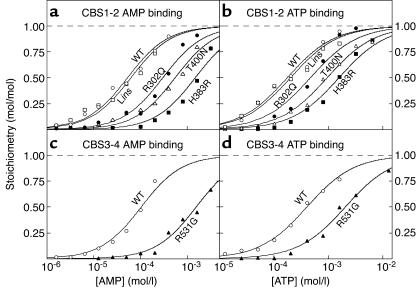
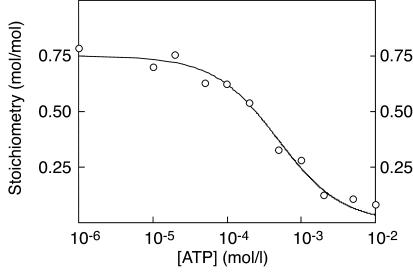
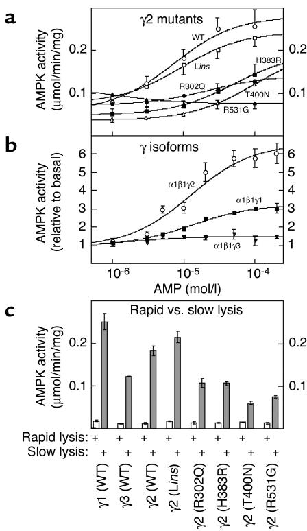
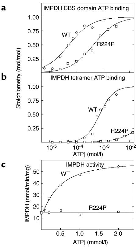
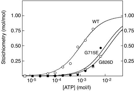
CBS domains are defined as sequence motifs that occur in several different proteins in all kingdoms of life. Although thought to be regulatory, their exact functions have been unknown. However, their importance was underlined by findings that mutations in conserved residues within them cause a variety of human hereditary diseases, including (with the gene mutated in parentheses): Wolff-Parkinson-White syndrome (gamma 2 subunit of AMP-activated protein kinase); retinitis pigmentosa (IMP dehydrogenase-1); congenital myotonia, idiopathic generalized epilepsy, hypercalciuric nephrolithiasis, and classic Bartter syndrome (CLC chloride channel family members); and homocystinuria (cystathionine beta-synthase). AMP-activated protein kinase is a sensor of cellular energy status that is activated by AMP and inhibited by ATP, but the location of the regulatory nucleotide-binding sites (which are prime targets for drugs to treat obesity and diabetes) was not characterized. We now show that tandem pairs of CBS domains from AMP-activated protein kinase, IMP dehydrogenase-2, the chloride channel CLC2, and cystathionine beta-synthase bind AMP, ATP, or S-adenosyl methionine,while mutations that cause hereditary diseases impair this binding. This shows that tandem pairs of CBS domains act, in most cases, as sensors of cellular energy status and, as such, represent a newly identified class of binding domain for adenosine derivatives.


Figures

Figure 1
Figure 1
Binding of AMP (a and c) or ATP (b and d) by the CBS1-2 construct (a and b) or the CBS3-4 construct (c and d). The fusion proteins were either the WT (open circles) or one of five point mutants (R302Q, filled circles; Lins, open squares; H383R, filled squares; T400N, open triangles; R531G, filled triangles). Data were fitted to a single-site binding model: bound = [nucleotide]/(Kd + [nucleotide]). The curves are theoretical curves obtained using the Kd values shown in Table 1.
Figure 2
Figure 2
Displacement of AMP from the CBS1-2 construct by ATP. A fixed concentration of [14C]AMP (180 μM) was incubated with the CBS1-2 construct in the presence of increasing concentrations of ATP. Data were fitted to the binding model: bound = AMP/[AMP + KdAMP(1 + [ATP]/KdATP)].
Figure 3
Figure 3
(a and b) Binding of AMP (a) and ATP (b) to the GST–CBS1-4 fusion protein from γ2 and WPWS mutants. (c and d) Binding of AMP (c) and ATP (d) by the GST–CBS1-4 fusion proteins from γ1, γ2, and γ3. The methodology was as for Figure 1, except that data were fitted to a two-site Hill plot model: bound = 2 × [nucleotide]h/(B0.5h + [nucleotide]h).
Figure 4
Figure 4
(a) Activation of recombinant α1β1γ2 heterotrimers, with or without WPWS mutations, by AMP. (b) Activation of recombinant α1β1γ1, α1β1γ2, and α1β1γ3 heterotrimers by AMP. (c) Activation of α1β1γ2 heterotrimers, with or without WPWS mutations, by slow versus rapid lysis. Plasmids expressing myc-tagged α1 and β1 plus one of the subunits γ1, γ2 (with or without WPWS mutations), and γ3 were expressed in CCL13 cells, and the recombinant complexes were immunoprecipitated using anti-myc antibodies. AMPK activity was then determined at various concentrations of AMP. In a and b, the cells were harvested by slow lysis to elicit maximal phosphorylation (32); in c, the cells were harvested by rapid or slow lysis and the assays were conducted at 200 μM AMP. Results are means ± SE for duplicate immunoprecipitations.
Figure 5
Figure 5
(a and b) Binding of ATP by GST fusions of the isolated CBS domain pair (residues 112–232) (a) and full-length IMPDH2 (residues 1–514) (b). (c) Activity of full-length IMPDH2 as a function of ATP concentration. Results were obtained for both the WT sequence and an R224P mutation. Data in a and b were fitted to a single-site binding model as for Figure 1. Data in c were fitted to the model: activity = basal + {[(stimulation × basal) – basal] × [ATP]h}/(A0.5h + [ATP]h).
Figure 6
Figure 6
Binding of ATP by a GST fusion of the isolated CBS domain pair (residues 582–840) from CLC2, and binding of ATP by G715E and G826D mutations. Data were fitted to a single-site binding model as for Figure 1.
Figure 7
Figure 7
Binding of SAM by a GST fusion of the isolated CBS domain pair (residues 416–551) from cystathionine β-synthase, and binding of SAM by a D444N mutation. Data were fitted to a single-site binding model as for Figure 1.
Figure 8
Figure 8
Model of the N-terminal pair of CBS domains (CBS1 and CBS2) from the γ2 subunit of AMPK. The picture is a view of a molecular-surface representation made using the program GRASP (Department of Biochemistry, Columbia University, New York, New York, USA) (51), with CBS1 on the left. The model was made using MODELLER 6 (Department of Biopharmaceutical Sciences, University of California San Francisco, San Francisco, California, USA) (52) and was based on the atomic coordinates of the CBS domain pair from a bacterial IMPDH (Protein Data Bank code 1ZFJ) (34). Electrostatic potential at the surface is depicted as red for negative and blue for positive. The approximate positions of residues mutated in WPWS are shown. R302 and H383 occupy equivalent positions in CBS1 and CBS2, respectively, and are adjacent to each other in the model. Part of residue T400 projects into the hydrophobic cleft between the two domains.

Comment in

References

    1. Bateman A. The structure of a domain common to archaebacteria and the homocystinuria disease protein. Trends Biochem. Sci. 1997;22:12–13. - PubMed
    1. Kluijtmans LA, et al. Defective cystathionine β-synthase regulation by S-adenosylmethionine in a partially pyridoxine responsive homocystinuria patient. J. Clin. Invest. 1996;98:285–289. - PMC - PubMed
    1. Bowne SJ, et al. Mutations in the inosine monophosphate dehydrogenase 1 gene (IMPDH1) cause the RP10 form of autosomal dominant retinitis pigmentosa. Hum. Mol. Genet. 2002;11:559–568. - PMC - PubMed
    1. Kennan A, et al. Identification of an IMPDH1 mutation in autosomal dominant retinitis pigmentosa (RP10) revealed following comparative microarray analysis of transcripts derived from retinas of wild-type and Rho(–/–) mice. Hum. Mol. Genet. 2002;11:547–557. - PubMed
    1. Pusch M. Myotonia caused by mutations in the muscle chloride channel gene CLCN1. Hum. Mutat. 2002;19:423–434. - PubMed

Publication types

MeSH terms